
Fig. 2
- ID
- ZDB-FIG-151119-6
- Publication
- Bhumika et al., 2015 - Decreased thyroid hormone signaling accelerates the reinnervation of the optic tectum following optic nerve crush in adult zebrafish
- Other Figures
- All Figure Page
- Back to All Figure Page
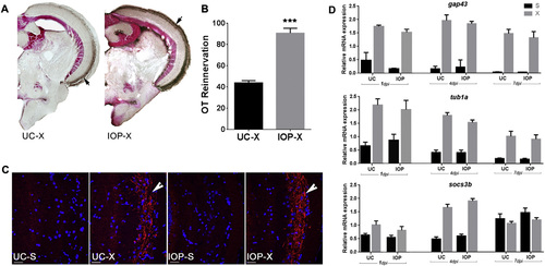
IOP treatment following ONC facilitates OT reinnervation. A. Reinnervation of the OT by RGC axons at 7 dpi in UC and IOP fish. Brown staining indicated with arrow represents biocytin labeled RGC axons. B. Quantification of the area covered by RGC axons in the OT of UC and IOP fish. Data represent mean ± SEM (n = 5 animals per group). ***p < 0.0001 by unpaired t-test. C. Immunofluorescent detection of Gap43 protein (red) in RGC axons (arrow) arriving in the OT. Cell nuclei are stained with DAPI (blue). Pictures are representative images for n = 5 animals/group. Scale bar 20 µm. D. Lowering intracellular TH availability did not affect the expression of regeneration associated genes in the retina. qPCR shows that the relative mRNA level of gap43, tuba1, and socs3b is significantly upregulated at 1, 4 and 7 dpi in both UC and IOP fish. Upregulation is however similar in both groups. Data represent mean ± SEM (n = 4 retina pools per group). For all panels: UC-untreated control, IOP—iopanoic acid treated, S—sham operated, X—optic nerve crushed, OT—optic tectum. |
Reprinted from Molecular and cellular neurosciences, 68, Bhumika, S., Lemmens, K., Vancamp, P., Moons, L., Darras, V.M., Decreased thyroid hormone signaling accelerates the reinnervation of the optic tectum following optic nerve crush in adult zebrafish, 92-102, Copyright (2015) with permission from Elsevier. Full text @ Mol. Cell Neurosci.