
Fig. 1
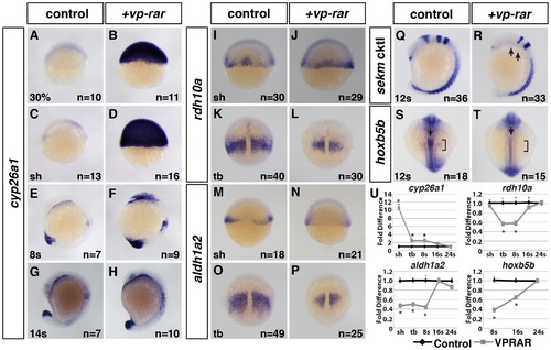
VP-RAR promotes a prolonged increase in cyp26a1 expression. (A–H) Wholemount ISH shows a dramatic increase in cyp26a1 expression in vp-rar mRNA injected embryos compared to WT siblings at 30% epiboly through 14 s stage. (I–P) ISH of RA synthesizing genes rdh10a and aldh1a2 shows modestly decreased expression in vp-rar mRNA injected embryos compared to WT at the shield stage (I,J,M,N), with dramatic decreases found by the tailbud stage (K,L,O,P). (Q,R) ISH using a six6b (anterior diencephalon), eng2a (midbrain–hindbrain boundary), egr2b (rhombomeres 3 and 5), and myod1 (adaxial cells and somites) cocktail (referred to here at sekm cktl) shows a lack of eng2a and severely reduced six6b and rhombomere 3 (anterior egr2b) in vp-rar mRNA injected compared to WT. Arrows in R indicate the loss of eng2a and reduced rhombomere 3. (S,T) Expression of hoxb5b is decreased in the lateral plate mesoderm (brackets) and anterior spinal cord (arrows) in vp-rar mRNA injected embryos compared to WT siblings. (U) RT-qPCR with cyp26a1, rdh10a, aldh1a2, and hoxb5b expression in vp-rar mRNA injected embryos at the shield (sh) through 24 s stages. Asterisks indicate statistically different from control (p<0.05) using Student′s t-test. |
Reprinted from Developmental Biology, 405(1), Rydeen, A., Voisin, N., D'Aniello, E., Ravisankar, P., Devignes, C.S., Waxman, J.S., Excessive feedback of Cyp26a1 promotes cell non-autonomous loss of retinoic acid signaling, 47-55, Copyright (2015) with permission from Elsevier. Full text @ Dev. Biol.