
FIGURE
Fig. S2
- ID
- ZDB-FIG-151002-45
- Publication
- Riemer et al., 2015 - A functional Bucky ball-GFP transgene visualizes germ plasm in living zebrafish
- Other Figures
- All Figure Page
- Back to All Figure Page
Fig. S2
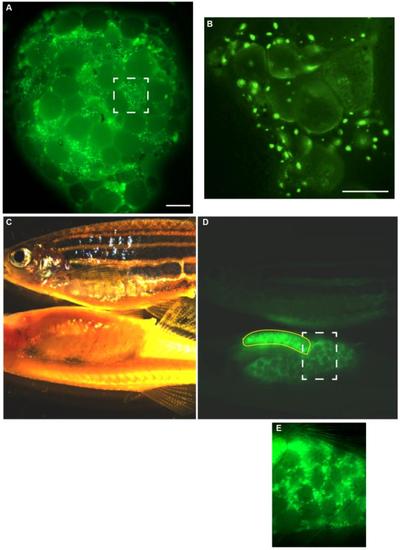
Expression of Buc-GFP in ovaries. (A, B) Living ovary from an adult F1- female transgenic for Buc-GFP. Dashed square highlights the magnified area in B. Scale bar: 700 µm. (B) Magnification showing that the fluorescence of Buc-GFP is obscured during oogenesis probably by the uptake of yolk protein. Scale bar: 200 µm. (C-E) Comparison of a wild-type (top) and casper female (bottom) carrying a Buc-GFP transgene in incident (C) and fluorescent light (D). Yellow line encompasses the stomach with autofluorescent food residues in the digestive tract. Dashed square highlights the magnified area in (E). |
Expression Data
Expression Detail
Antibody Labeling
Phenotype Data
Phenotype Detail
Acknowledgments
This image is the copyrighted work of the attributed author or publisher, and
ZFIN has permission only to display this image to its users.
Additional permissions should be obtained from the applicable author or publisher of the image.
Reprinted from Gene expression patterns : GEP, 18(1-2), Riemer, S., Bontems, F., Krishnakumar, P., Gömann, J., Dosch, R., A functional Bucky ball-GFP transgene visualizes germ plasm in living zebrafish, 44-52, Copyright (2015) with permission from Elsevier. Full text @ Gene Expr. Patterns