
Fig. S4
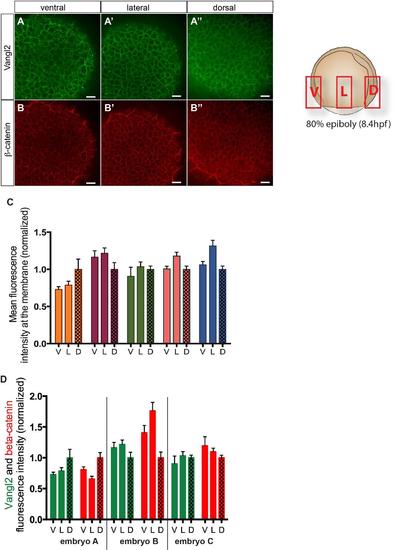
Vangl2 membrane localization in ventral, lateral or dorsal domain of the embryo does not show domain specific preference. (A-B′′) Confocal images acquired in the ventral (A,B), lateral (A′B′) and dorsal (A′′,B′′) domains in the embryo. Images were taken in all 3 domains of the same embryo and analyzed separately. Embryos at 80% epiboly stage (8.4hpf) were analyzed. (C) Quantitative data obtained from the analysis of the Vangl2 staining fluorescence intensity at the cell membrane in 5 different embryos (each embryo’s D, V, L data set is in different color on the histogram). (D) Quantitative data comparing β-catenin (control) and Vangl2 staining for 3 different embryos in all D, L V domains, showing no domain specific preference for neither protein. All data were normalized such as dorsal=1. Scale bars, 20 µm. |