
Fig. 2
- ID
- ZDB-FIG-150923-2
- Publication
- Helmprobst et al., 2015 - Presynaptic architecture of the larval zebrafish neuromuscular junction
- Other Figures
- All Figure Page
- Back to All Figure Page
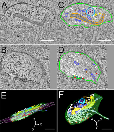
Three-dimensional model of an NMJ of a 4-dpf zebrafish larva. Three serial tomograms (~600 nm overall thickness) were joined to show the extent of an NMJ. A and B show single slices of the tomograms. The tomograms were annotated in 3dmod (C,D) to highlight cellular structures in the synapse. The synapse is next to a muscle cell (M) with a synaptic cleft (SC) between and surrounded by a membrane (light green). In the synapse, synaptic vesicles (white), mitochondria (orange), endoplasmic reticulum (light blue), two types of dense-core vesicles (brown and yellow), and large multivesicular bodies (turquois) with internal vesicles (purple) were found. Docked synaptic vesicles at the presynaptic membrane are annotated in red. At the active zone we observed electron-dense structures (green), which are surrounded by many docked vesicles. E and F show the whole 3D model of the synapse. The magenta planes in E show the borders, where the three tomograms were joined. Scale bars = 500 nm. |