
Fig. S2
- ID
- ZDB-FIG-150918-20
- Publication
- Rossi et al., 2015 - The SLC7A7 Transporter Identifies Microglial Precursors prior to Entry into the Brain
- Other Figures
- All Figure Page
- Back to All Figure Page
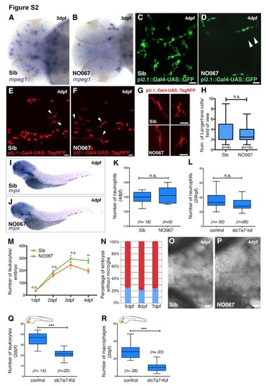
The NO067 phenotype is specific for microglia. Related to Figure 2. (A and B) Dorsal view of 3 dpf sibling (A) and NO067 mutant brain (B) stained with an antisense mpeg1 RNA probe. (C and D) Dorsal view of a representative pU.1::GFP sibling (C) and NO067 mutant (D) brain at 4 dpf . NO067 lacks microglia but retains circulating monocytes (white arrowheads).(E and F) Representative region of the superficial layers of the trunk in a sibling (E) and a NO067 mutant (F) at 4 dpf. White arrowheads indicate Langerhans cells expressing TagRFP (pU.1:: TagRFP). (G) Representative Langerhans cells in a 4 dpf embryo. There are no morphological differences between sibling cells (top panels) and mutant cells (bottom panels). (H) Quantifications of the number of Langerhans cells within the region of interest (as shown in E and F). (I and J) WISH for mpx in a sibling (I) and a NO067 mutant (J) embryo at 4 dpf (side view). (K) Quantifications of the number of neutrophils (mpx) in siblings and mutants at 4 dpf. (L) Quantifications of the number of neutrophils (mpx) in wild type and morphants at 24 hpf. (M) Quantifications of the number of leukocytes in siblings (green line) and NO067 homozygous mutants (orange line). **p<0.005. Mann-Whitney test. (N) Percentage of embryos lacking microglia (blue bars) in clutches derived from crossing NO067 heterozygotes parents. Error bars show SEM. (O and P) Representative examples of sibling (O) and mutant (P) brains at 4 dpf (brightfield, dorsal view). In the absence of microglia apoptotic corpses accumulate in the mutant brain. (Q and R) Quantifications of the number of leukocytes (Q) and macrophages (R) in the head of wild-type and slc7a7 morphant embryo. ***p<0.0005. Mann-Whitney test. Scale bars for all images: 25 μm. Sib, sibling. n=num. of embryos. |