
Fig. 4
- ID
- ZDB-FIG-150617-4
- Publication
- Wu et al., 2015 - Hypoxia-Induced Retinal Neovascularization in Zebrafish Embryos: A Potential Model of Retinopathy of Prematurity
- Other Figures
- All Figure Page
- Back to All Figure Page
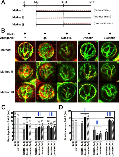
VEGF inhibitors rescue overangiogenesis and leakage in the CoCl2-induced hypoxic retinal vasculature. (A) Three conditions were designed to mimic the clinical situation and to examine the effects of treatment. Method I: Tg(fli1a:EGFP) embryos were treated with CoCl2 (black line) and the inhibitor (red dotted line) simultaneously from 1 dpf to 5 dpf. Method II: Prior to CoCl2 treatment (black line) at 3 dpf, we immersed embryos in the inhibitor (red dotted line) for 2 days. Method III: Following CoCl2treatment, the inhibitor was added to the solution at 3 dpf. All embryos were injected with TAMRA dye at 4 dpf, and their retinal vasculatures were subsequently observed at 5 dpf. (B) CoCl2-treated embryos showed TAMRA dye seepage (red) from the intraocular vessels (green). Similar leakages and excessive neovascularization were appeared in the control IgG-treated groups (CoCl2 and normal human control IgG cotreatment). Three candidate inhibitors, SU5416, bevacizumab, and ranibizumab, reversed the effect of CoCl2 on retinal vessels. (C) Statistical analyses of branch points showed significant rescue in inhibitor-treated embryos. The total numbers of embryos for analyses are indicated on the top of each bar. (D) However, the survival rates (four independent experiments, n = 35 in each) showed low toxicity under the Method I condition. Each bar represents the mean ± SEM of * (p < 0.0125) compared with the CoCl2 = 5 mM group. |
| Gene: | |
|---|---|
| Fish: | |
| Condition: | |
| Anatomical Term: | |
| Stage: | Day 5 |