
Fig. 3
- ID
- ZDB-FIG-150617-3
- Publication
- Wu et al., 2015 - Hypoxia-Induced Retinal Neovascularization in Zebrafish Embryos: A Potential Model of Retinopathy of Prematurity
- Other Figures
- All Figure Page
- Back to All Figure Page
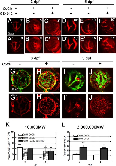
Leakage analyses of CoCl2-treated retinal vasculature performed using two types of fluorescent dyes: 10,000 MW dextran, and 2,000,000 MW TAMRA. (A–F′) Injected dextran (red) in the vessels became obscured, with apparent dextran leakage within 9 and 15 minutes in treated zebrafish embryos 3 and 5 dpf, respectively (B′, C′, E′, F′); however, in the control embryos, leakage was observed in the retinal vessels (A′). No leakage was apparent in the normal tight endothelium of 5-dpf embryos (D′). (G–J′) Upon injection, most of the TAMRA dye (red) was contained in the vasculature, and leakage was observed 24 hours later. Scant leakage occurred at the center of the control retina (G, I), whereas apparent leakage was observed in CoCl2-treated vasculatures (H, J). (K, L) The fluorescence values in 3- and 5-dpf embryos were quantified using Image J to show the dynamic changes of the 10,000 MW and 2,000,000 MW dyes. Data are presented as the mean ± standard deviation from three to five embryos. |
| Gene: | |
|---|---|
| Fish: | |
| Condition: | |
| Anatomical Term: | |
| Stage Range: | Protruding-mouth to Day 5 |
| Fish: | |
|---|---|
| Conditions: | |
| Observed In: | |
| Stage Range: | Protruding-mouth to Day 5 |