
Fig. 5
- ID
- ZDB-FIG-150602-22
- Publication
- Liu et al., 2015 - Differential expression of protocadherin-19, protocadherin-17, and cadherin-6 in adult zebrafish brain
- Other Figures
- All Figure Page
- Back to All Figure Page
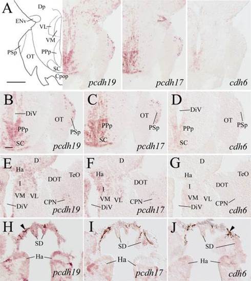
Expression of pcdh19, pcdh17, and cdh6 in the posterior preoptic areas, epithalamus, ventral thalamus, and pretectum. Top panel images show low magnifications of the brain from adjacent sections at a level indicated in Fig. 1. B–D: Higher magnifications of the posterior preoptic areas and the suprachiasmatic nucleus (SC) shown in the top panels. The remaining panels are from sections 100–150 µm posterior to those shown in B–D, showing epithalamus and ventral thalamus. Arrowheads in H–J indicate pigmented epithelium. For other abbreviations, see list. Scale bar = 200 µm for the top panels, 50 µm for the lower panels (B–J have the same magnification). |
| Genes: | |
|---|---|
| Fish: | |
| Anatomical Terms: | |
| Stage: | Adult |