
Fig. 3
- ID
- ZDB-FIG-150526-9
- Publication
- Petersen et al., 2015 - The adhesion GPCR GPR126 has distinct, domain-dependent functions in Schwann cell development mediated by interaction with laminin-211
- Other Figures
- All Figure Page
- Back to All Figure Page
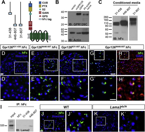
GPR126-NTF Binds SC-Derived Lama2 (A) Diagram showing domains in human GPR126-NTF and fusion constructs for three different GPR126-NTF fragments. (B) Detection of GPR126 cleavage in vitro and in vivo. P6 mouse sciatic nerve and 293T cells transfected with either mouse Gpr126 cDNA or empty vector were lysed and immunoblotted with rabbit anti-GPR126 CTF antibody. A specific 35 kDa GPR126 CTF was detected in Gpr126-transfected cells and in P6 mouse sciatic nerve. IB, immunoblotting. (C) Fusion constructs were transfected into HEK293T cells to generate fusion recombinant proteins. Purified proteins were verified by western blot. Two bands were detected in GPR126N31–807 transfected media due to the second cleavage between aa 440 and 441; the higher band is full-length NTF. (D–F) Putative ligand binding on P3 sciatic nerves. Anti-hFc immunostaining is green and nuclear counterstain is Hoechst 33342 (blue). Magnification of boxed regions in (D)–(F) (the scale bar represents 20 µm) is indicated in (D′)–(F′) (the scale bar represents 10 µm). (D) No binding was detected with the GPR126N31–438 hFc fragment. (E and F) The GPR126N446–807 (E) and full-length GPR126N31–807 hFc (F) fragments reveal the same ligand binding pattern. (G and H) The GPR126N446–807 hFc fragment (green) colocalizes with SCs marked by S100β (red) in P10 sciatic nerves. Nuclear counterstain is 42,6-diamidino-2-phenylindole (DAPI) (blue). The scale bars represent 20 µm (G and H) and 10 µm (G′ and H′). (I) GPR126N446–807 binds Lama2 in vivo. GPR126N446–807 and GPR126N31–438 proteins were mixed with P3 sciatic nerve lysate and immunoprecipitated with anti-hFc antibodies. Lama2 specifically coimmunoprecipitates with GPR126N446–807. Input: 15 s exposure; Lama2, 1 min exposure. IP, immunoprecipitation. (J and K) Putative ligand binding performed on P10 sciatic nerves of WT and Lama2dy3k/dy3k mice. Anti-hFc immunostaining is green and nuclear counterstain is Hoechst 33342 (blue). Magnification of boxed regions in (J) and (K) (the scale bar represents 10 µm) is indicated in (J′) and (K′) (the scale bar represents 2.7 µm). GPR126N446–807 binding is robust in WT sciatic nerve (J) but not detected in Lama2dy3k mutant sciatic nerve (K). |