
Fig. 7
- ID
- ZDB-FIG-150428-36
- Publication
- Guzman et al., 2013 - A stem cell proliferation burst forms new layers of P63 expressing suprabasal cells during zebrafish postembryonic epidermal development
- Other Figures
- All Figure Page
- Back to All Figure Page
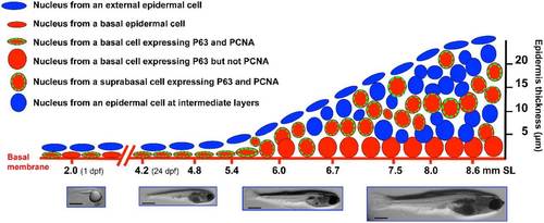
Model for epidermal stratification during postembryonic development in zebrafish. This diagram shows the labeling in the nuclei from epidermal cells, through different postembryonic developmental stages, as was observed in this work. By 1 dpf the embryo has an approximate size of 2mm in length, the epidermis is formed by two cell layers, the external layer and the basal layer. The basal layer is known to express the P63 transcription factor (a known marker for epidermal stem cells). Just before epidermal stratification initiates (SL 4.2–4.8mm) there are high levels of proliferation in the basal layer. When the fish reach a size of SL 5.4–6mm, epidermal stratifications begins. At this point the nuclei of basal cells became rounded and these cells reduce proliferation. A new layer of suprabasal cells appears which also express P63 and shows an active cell proliferation. At stages of SL 8.0–8.6 the epidermis is 5–8 layers thick (depending of the body region) and suprabasal cells are still proliferative while basal cells have completely stop proliferation. It is possible that suprabasal cells are “transient-amplifying cells”. Bars are 1mm in length. |