
- Title
-
A stem cell proliferation burst forms new layers of P63 expressing suprabasal cells during zebrafish postembryonic epidermal development
- Authors
- Guzman, A., Ramos-Balderas, J.L., Carrillo-Rosas, S., and Maldonado, E.
- Source
- Full text @ Biol. Open
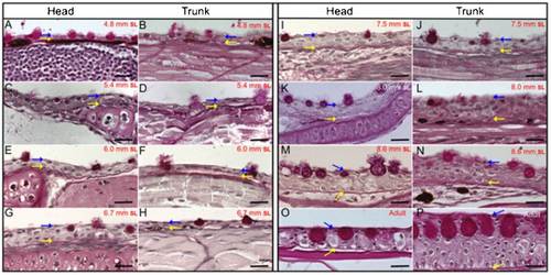
Histological longitudinal sections and periodic acid-Schiff staining of juvenile fish with a SL of 4.8mm to 8.6mm and from adult zebrafish. (A, C, E, G, I, K, M and O) Sections from the head region. (B, D, F, H, J, L, N and P) Sections from the dorsal trunk area. (A, B), (C, D), (E, F), (G, H), (I, J), (K, L) and (M, N) are from fish with a SL of 4.8mm, 5.4mm, 6mm, 6.7mm, 7.5mm, 8mm and 8.6mm, respectively. (O, P) Sections of the head and dorsal trunk from adult fish. Blue arrows label the topmost limit of the epidermis and yellow arrows label the basal membrane, which is specifically labeled by PAS. (C–F) Epidermal stratification and thickening was initiated when larvae attained a SL of 5.4 and 6mm. (G–N) When larvae reached a SL of 6.7–8. 6mm, stratification notably thickened the epidermis. During this time, the mucous cells undergo a considerable increase in size, which might give the impression that these images were not taken at the same magnification; however, all images shown here were obtained with a 63× oil objective (Zeiss). The large spaces visible between the skin and other tissues may be artifactual. Bars are 20µm in length. |
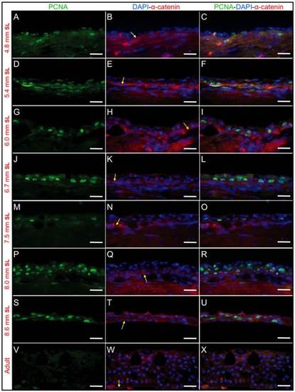
PCNA and α-catenin immunofluorescence staining reveals cell proliferation during epidermal stratification in the dorsal trunk area. Anti-PCNA labels proliferating cells, while anti-α-catenin labels both the epidermis and dermis but is concentrated in the basal membrane, which allows the discrimination of these two regions. (A-C), (D-F), (G-I), (J-L), (M-O), (P-R) and (S-U) show immunofluorescence staining from longitudinal sections of larvae with a SL of 4.8mm, 5.4mm, 6mm, 6.7mm, 7.5mm, 8mm and 8.6mm SL, respectively. PCNA labeling is observed specifically in basal layer cells from SL 4.8-5.4mm larvae (A-F), but later is observed in suprabasal layers (compare C, L and R). (V-X) are immunostained longitudinal sections from adult fish; no PCNA staining can be observed in this representative region. In some samples (like S-U) the epidermis get separated from the dermis but this is artifactual. Yellow arrows mark the epidermal basal membrane where α-catenin labeling concentrates. Bar length is 20 µm. |
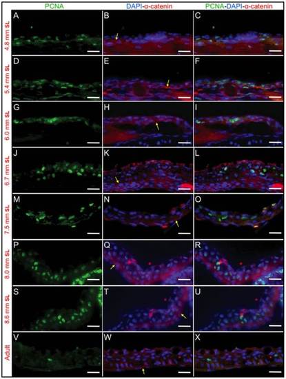
PCNA and α-catenin immunostaining to show cell proliferation during epidermal stratification in the head area. Anti-PCNA labels cells in proliferation and anti-α-catenin labels the basal membrane that separates the epidermis and dermis regions. (A–C), (D–F), (G–I), (J–L), (M–O), (P–R) and (S–U) show immunostained longitudinal sections of larvae with a SL of 4.8mm, 5.4mm, 6mm, 6.7mm, 7.5mm, 8mm and 8.6mm, respectively. (V–X) show immunostained longitudinal sections in adult fish. (A, D, G, J, M, P, S and V) PCNA immunostaining. (B, E, H, K, N, Q, T and W) Anti-α-catenin immunofluorescence merged with DAPI staining to label the nuclei. (C, F, I, L, O, R, U and X) Merged images of PCNA, α-catenin and DAPI staining. In some samples (like M–O) the epidermis get separated from the dermis, however this is artifactual. Yellow arrows mark the epidermal basal membrane where α-catenin labeling is abundant. Bar length is 20 µm. |
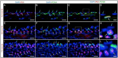
Zebrafish epidermal stratification as a consequence of the proliferation of P63-expressing cells. (A–D) Longitudinal sections from larvae with a SL of 4.2mm. (E–H) Longitudinal sections of larvae with a SL of 6mm. (I–L) Longitudinal sections of larvae with a SL of 7.5mm. (A, E and I) P63 and DAPI labeling. (B, F and J) PCNA and DAPI immunofluorescence. (C, G and K) Merged P63, PCNA and DAPI stainings; the colocalization of P63 and DAPI appears magenta, and the colocalization of P63 and PCNA appears yellow. (D, H and L) Higher magnification images of the regions marked by squares in (C, G and H), respectively. Prior to the initiation of stratification (SL of 4.2mm), basal cells (P63 positive) express the proliferation marker PCNA; later, for a SL of 6mm, new suprabasal cells appear, also exhibiting P63 and PCNA co-labeling. For a SL of 7.5mm SL, suprabasal cells (P63 positive) are abundant, and only suprabasal cells show PCNA labeling. Blue arrows label cells from the external epidermal layer, yellow arrows mark suprabasal cells at intermediate layers that are positive for PCNA and P63 expression, white arrows label P63 positive cells from the basal layer. Bars are 20µm in length, except in (D, H and L), where they are 5µm in length. |
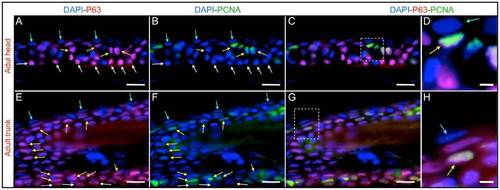
PCNA and P63 immunofluorescence in the adult zebrafish epidermis. (A–D) Head region. (E–H) Dorsal trunk area, in a region that contains only scales. (A and E) DAPI and P63 labeling. (B and F) DAPI and PCNA immunofluorescence. (C and G) Merged P63, PCNA and DAPI immunostainings. (D and H) Higher magnification images of the regions marked by squares in (C and G), respectively. Suprabasal P63-positive cells persist in adulthood and remain actively proliferating. Blue arrows mark cells from the external epidermal layer, yellow arrows label suprabasal cells at intermediate layers that are positive for PCNA and P63 expression, white arrows mark P63 positive cells from the basal layer. Bars are 20µm in length, except in (D, H and L) where they are 5µm in length. |
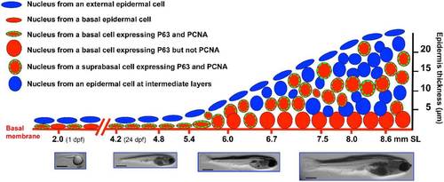
Model for epidermal stratification during postembryonic development in zebrafish. This diagram shows the labeling in the nuclei from epidermal cells, through different postembryonic developmental stages, as was observed in this work. By 1 dpf the embryo has an approximate size of 2mm in length, the epidermis is formed by two cell layers, the external layer and the basal layer. The basal layer is known to express the P63 transcription factor (a known marker for epidermal stem cells). Just before epidermal stratification initiates (SL 4.2–4.8mm) there are high levels of proliferation in the basal layer. When the fish reach a size of SL 5.4–6mm, epidermal stratifications begins. At this point the nuclei of basal cells became rounded and these cells reduce proliferation. A new layer of suprabasal cells appears which also express P63 and shows an active cell proliferation. At stages of SL 8.0–8.6 the epidermis is 5–8 layers thick (depending of the body region) and suprabasal cells are still proliferative while basal cells have completely stop proliferation. It is possible that suprabasal cells are “transient-amplifying cells”. Bars are 1mm in length. |