
Fig. 2
- ID
- ZDB-FIG-150428-31
- Publication
- Guzman et al., 2013 - A stem cell proliferation burst forms new layers of P63 expressing suprabasal cells during zebrafish postembryonic epidermal development
- Other Figures
- All Figure Page
- Back to All Figure Page
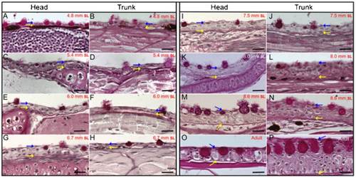
Histological longitudinal sections and periodic acid-Schiff staining of juvenile fish with a SL of 4.8mm to 8.6mm and from adult zebrafish. (A, C, E, G, I, K, M and O) Sections from the head region. (B, D, F, H, J, L, N and P) Sections from the dorsal trunk area. (A, B), (C, D), (E, F), (G, H), (I, J), (K, L) and (M, N) are from fish with a SL of 4.8mm, 5.4mm, 6mm, 6.7mm, 7.5mm, 8mm and 8.6mm, respectively. (O, P) Sections of the head and dorsal trunk from adult fish. Blue arrows label the topmost limit of the epidermis and yellow arrows label the basal membrane, which is specifically labeled by PAS. (C–F) Epidermal stratification and thickening was initiated when larvae attained a SL of 5.4 and 6mm. (G–N) When larvae reached a SL of 6.7–8. 6mm, stratification notably thickened the epidermis. During this time, the mucous cells undergo a considerable increase in size, which might give the impression that these images were not taken at the same magnification; however, all images shown here were obtained with a 63× oil objective (Zeiss). The large spaces visible between the skin and other tissues may be artifactual. Bars are 20µm in length. |