
Fig. S5
- ID
- ZDB-FIG-150422-69
- Publication
- Olena et al., 2015 - miR-216a regulates snx5, a novel notch signaling pathway component, during zebrafish retinal development
- Other Figures
- All Figure Page
- Back to All Figure Page
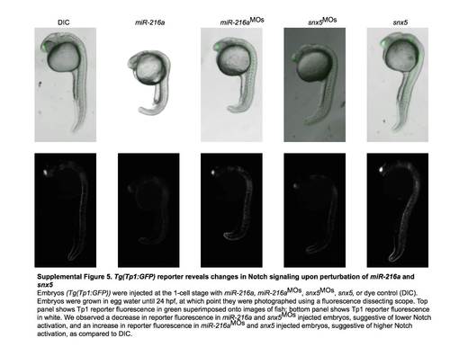
Tg(Tp1:GFP) reporter reveals changes in Notch signaling upon perturbation of miR-216a and snx5. Embryos (Tg(Tp1:GFP)) were injected at the 1-cell stage with miR-216a, miR-216aMOs, snx5MOs, snx5, or dye control (DIC). Embryos were grown in egg water until 24 hpf, at which point they were photographed using a fluorescence dissecting scope. Top panel shows Tp1 reporter fluorescence in green superimposed onto images of fish; bottom panel shows Tp1 reporter fluorescence in white. We observed a decrease in reporter fluorescence in miR-216a and snx5MOs injected embryos, suggestive of lower Notch activation, and an increase in reporter fluorescence in miR-261aMOs and snx5 injected embryos, suggestive of higher Notch activation, as compared to DIC. |
Reprinted from Developmental Biology, 400(1), Olena, A.F., Rao, M.B., Thatcher, E.J., Wu, S.Y., Patton, J.G., miR-216a regulates snx5, a novel notch signaling pathway component, during zebrafish retinal development, 72-81, Copyright (2015) with permission from Elsevier. Full text @ Dev. Biol.