
Fig. 2
- ID
- ZDB-FIG-150422-61
- Publication
- Olena et al., 2015 - miR-216a regulates snx5, a novel notch signaling pathway component, during zebrafish retinal development
- Other Figures
- All Figure Page
- Back to All Figure Page
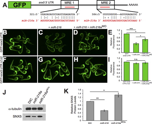
snx5 is a target of miR-216a. The coding sequence of GFP was fused to the 32UTR of snx5. Predicted pairing of each MRE in the 32 UTR (black) and miR-216a (red) are pictured. (B) Embryos injected at the 1-cell stage with GFP-snx5 32 UTR reporter mRNA alone, with miR-216a (C), or with miR-216a and miR-216aMO (D) were imaged using a fluorescence dissecting scope at 1 dpf. (F) Both MREs were deleted from the GFP-snx5 32 UTR reporter. Embryos injected at the 1-cell stage with this mRNA alone, with miR-216a (G), or with miR-216a and miR-216aMO (H) were imaged at 1 dpf using a fluorescence dissecting scope. (E, I) Relative fluorescence was quantified using ImageJ, and comparisons were made using one-way ANOVA with Bonferroniós correction. (J) Western blots for SNX5 and alpha-tubulin were performed on protein lysates from 1 dpf zebrafish injected at 1-cell stage with dye control (DIC), miR-216a, or miR-216aMOs. (K) Band density was quantified using ImageJ, and comparisons were made using one-way ANOVA with Bonferroniós correction. *, p<0.05; **, p<0.01; ***, p<0.001. Error bars show SEM. |
Reprinted from Developmental Biology, 400(1), Olena, A.F., Rao, M.B., Thatcher, E.J., Wu, S.Y., Patton, J.G., miR-216a regulates snx5, a novel notch signaling pathway component, during zebrafish retinal development, 72-81, Copyright (2015) with permission from Elsevier. Full text @ Dev. Biol.