
Fig. 6
- ID
- ZDB-FIG-150408-22
- Publication
- Strzyz et al., 2015 - Interkinetic Nuclear Migration Is Centrosome Independent and Ensures Apical Cell Division to Maintain Tissue Integrity
- Other Figures
- All Figure Page
- Back to All Figure Page
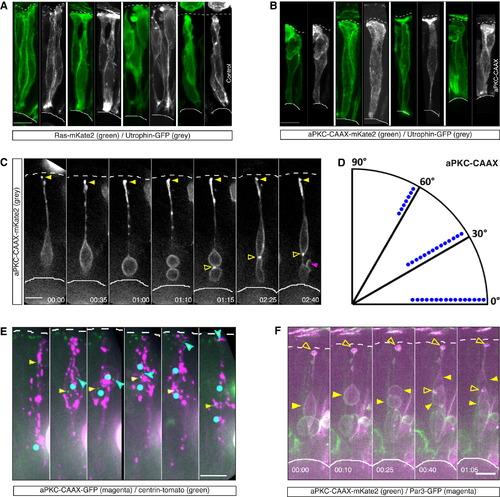
Interference with Actin Distribution via Membranous Expression of aPKC-CAAX Leads to Nonapical Divisions (A) Confocal scans of actin organization in control cells. Cells were coinjected with HS-Ras-mKate2 (green, membrane) and HS-Utrophin-GFP (F-actin, gray). Actin is organized in distinct filaments. HS was performed 7 hr prior to fixation. (B) Confocal scans of actin organization in HS-aPKC-CAAX-mKate2 (green) expressing cells coinjected with HS-Utrophin-GFP (F-actin, gray). Actin is diffusely organized without a clear filamentous structure along the cell membrane, despite a clear apical signal. HS is 7 hr prior to fixation. (C) Time-lapse of a cell expressing aPKC-CAAX-mKate2 (gray). The cell divides nonapically with a horizontal cleavage plane (01:10), while maintaining its apical attachment (yellow filled arrow). The more apical daughter cell maintains its apical process attachment, while the more basal daughter forms an ectopic connection to the sister cell (open arrow). Later, the basal daughter shows protrusive activity (magenta arrow). HS was performed 9.5 hr prior to time-lapse. Time is in hr:min. The frames are from Movie S7. See also Figure S3. (D) Division angles of the aPKC-CAAX expressing cells dividing nonapically. Dots indicate individual cells. Cells lose clear bias for perpendicular divisions (angles 60° to 90°) observed in controls (Figure 5B), n = 37 cells, 8 embryos. (E) Time-lapse of a cell expressing HS-aPKC-CAAX-GFP (magenta) and centrin-tomato (green). The cell divides nonapically with a nonperpendicular cleavage plane (02:35). The centrosome of the more apical daughter (cyan-outlined arrow) descends to the apical surface. The centrosome of the more basal daughter (yellow arrow) remains basally. Blue dots represent soma/nuclear position. HS was performed 11.5 hr prior to time-lapse. Time is in hr:min. The frames are from Movie S7. See also Figure S3. (F) Time-lapse of a cell expressing HS-aPKC-CAAX-mKate2 (green) and HS-Par3-GFP (magenta). The cell divides nonapically with a horizontal cleavage plane (00:25), while maintaining apical process and apical Par3 signal (open arrow). Later, the basal daughter forms an ectopic, basal Par3 domain (open arrow). HS was performed 8 hr prior to the time-lapse. Time is in hr:min. The frames are from Movie S7. Scale bars represent 10 µm. The dotted line represents the apical surface, and the solid line represents the basal side. |
Reprinted from Developmental Cell, 32(2), Strzyz, P.J., Lee, H.O., Sidhaye, J., Weber, I.P., Leung, L.C., Norden, C., Interkinetic Nuclear Migration Is Centrosome Independent and Ensures Apical Cell Division to Maintain Tissue Integrity, 203-19, Copyright (2015) with permission from Elsevier. Full text @ Dev. Cell