
Fig. 1
- ID
- ZDB-FIG-150408-17
- Publication
- Strzyz et al., 2015 - Interkinetic Nuclear Migration Is Centrosome Independent and Ensures Apical Cell Division to Maintain Tissue Integrity
- Other Figures
- All Figure Page
- Back to All Figure Page
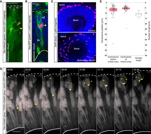
Mitotic Entry Is Not Restricted to the Apical Surface in Zebrafish Retinal NE; Centrosomes Are Maintained Apically throughout the Cell Cycle (A) Confocal scan of a cell expressing Ras-mKate2 (green) and centrin-GFP (magenta) in an embryo treated with 200 µM blebbistatin 1.5 hr before imaging. The cell shows nonapical mitotic rounding (filled arrow) and is associated with centrosomes (open arrows). (B) Confocal scan of a cell expressing Ras-GFP (green) stained for pH3 (magenta) in an embryo treated with 200 µM blebbistatin 1.5 hr before fixing. The cell features nonapical pH3 signal (filled arrow) while maintaining apical and basal attachments (open arrows). (C) Confocal scans of the retinae of an embryo treated with 200 µM blebbistatin 1.5 hr before fixing (lower) and a control embryo (upper) stained for pH3 (magenta). Blebbistatin treated cells enter mitosis nonapically (lower, arrows). (D) Time-lapse of the dynamics of the nucleus-centrosome pair with respect to cell cycle progression. PCNA-RFP labels nuclei and marks the cell cycle stage (gray). Ras-mKate2 (gray) labels cell membranes. Centrin-GFP (red) labels centrosomes. One nucleus is labeled with a yellow dot. The arrow highlights the position of the centrosome. Time is in hr:min. The frames are from Movie S1. (E) Centrosome position prior to centrosome splitting (left) and the mean value of the position of centrosomes at mitosis (middle) with respect to nuclear length (right), n = 51 cells, 4 embryos. Scale bars represent 10 µm. The dotted line represents the apical surface, and the solid line represents the basal side. |
Reprinted from Developmental Cell, 32(2), Strzyz, P.J., Lee, H.O., Sidhaye, J., Weber, I.P., Leung, L.C., Norden, C., Interkinetic Nuclear Migration Is Centrosome Independent and Ensures Apical Cell Division to Maintain Tissue Integrity, 203-19, Copyright (2015) with permission from Elsevier. Full text @ Dev. Cell