
Fig. S4
- ID
- ZDB-FIG-150331-35
- Publication
- Phng et al., 2015 - Formin-mediated actin polymerization at endothelial junctions is required for vessel lumen formation and stabilization
- Other Figures
- All Figure Page
- Back to All Figure Page
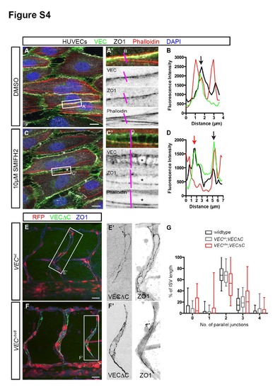
Junctional F-actin strengthens junction stability. Related to Figure 4. (A - D) HUVECs treated with either DMSO or 10µM SMIFH2 under high shear stress. In DMSO-treated cells, junctions at the long axis of cells exhibit linear morphology (A′) that consists of VEC, ZO1 and F-actin (black arrow, B). In SMIFH2-treated cells, intercellular gaps (*, C) appear between cells. Such junctions are characterized by a wider distribution of VEC, two ZO-1 lines and a loss of F-actin in some junctions (red arrow, D). Scale bar, 10µm. (E - G) ISV junction organization in 53hpf VECwt; Tg(fli1ep:GAL4FF)ubs3;Tg(UAS:mRFP);Tg(UAS:VECΔC-EGFP)ubs12 and VECubs8; Tg(fli1ep:GAL4FF)ubs3;Tg(UAS:mRFP);Tg(UAS:VECΔC-EGFP)ubs12 embryos. Embryos were immunostained with ZO1 antibody. The number of parallel junctions along the length of ISVs expressing VECΔC-EGFP was quantified (G). Scale bar, 20µm. |
Reprinted from Developmental Cell, 32, Phng, L.K., Gebala, V., Bentley, K., Philippides, A., Wacker, A., Mathivet, T., Sauteur, L., Stanchi, F., Belting, H.G., Affolter, M., Gerhardt, H., Formin-mediated actin polymerization at endothelial junctions is required for vessel lumen formation and stabilization, 123-32, Copyright (2015) with permission from Elsevier. Full text @ Dev. Cell