
|
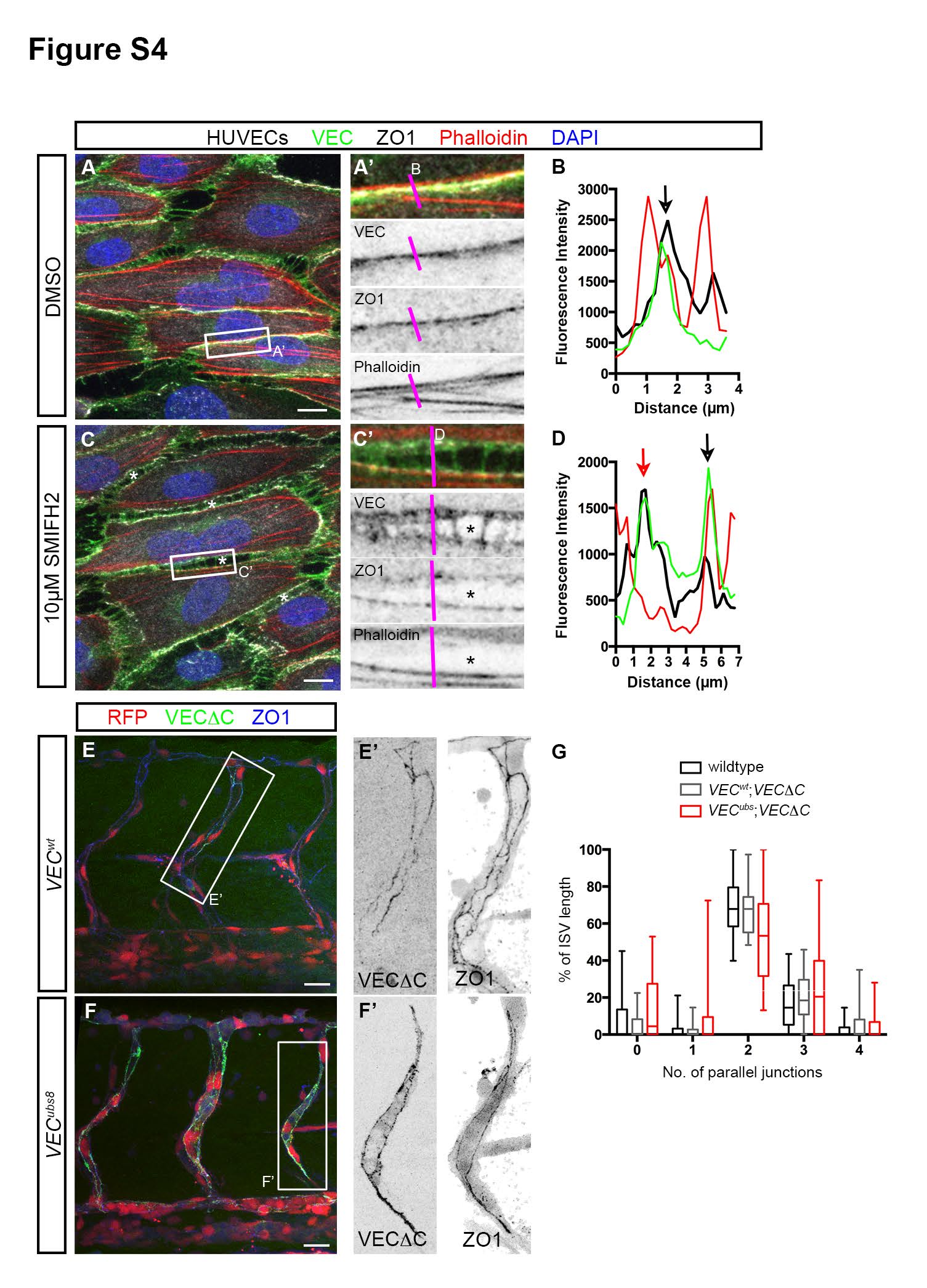
Fig. S4 Junctional F-actin strengthens junction stability. Related to Figure 4. (A - D) HUVECs treated with either DMSO or 10µM SMIFH2 under high shear stress. In DMSO-treated cells, junctions at the long axis of cells exhibit linear morphology (A′) that consists of VEC, ZO1 and F-actin (black arrow, B). In SMIFH2-treated cells, intercellular gaps (*, C) appear between cells. Such junctions are characterized by a wider distribution of VEC, two ZO-1 lines and a loss of F-actin in some junctions (red arrow, D). Scale bar, 10µm. (E - G) ISV junction organization in 53hpf VECwt; Tg(fli1ep:GAL4FF)ubs3;Tg(UAS:mRFP);Tg(UAS:VECΔC-EGFP)ubs12 and VECubs8; Tg(fli1ep:GAL4FF)ubs3;Tg(UAS:mRFP);Tg(UAS:VECΔC-EGFP)ubs12 embryos. Embryos were immunostained with ZO1 antibody. The number of parallel junctions along the length of ISVs expressing VECΔC-EGFP was quantified (G). Scale bar, 20µm.
Reprinted from Developmental Cell, 32, Phng, L.K., Gebala, V., Bentley, K., Philippides, A., Wacker, A., Mathivet, T., Sauteur, L., Stanchi, F., Belting, H.G., Affolter, M., Gerhardt, H., Formin-mediated actin polymerization at endothelial junctions is required for vessel lumen formation and stabilization, 123-32, Copyright (2015) with permission from Elsevier. Full text @ Dev. Cell