
Fig. 3
- ID
- ZDB-FIG-150331-30
- Publication
- Phng et al., 2015 - Formin-mediated actin polymerization at endothelial junctions is required for vessel lumen formation and stabilization
- Other Figures
- All Figure Page
- Back to All Figure Page
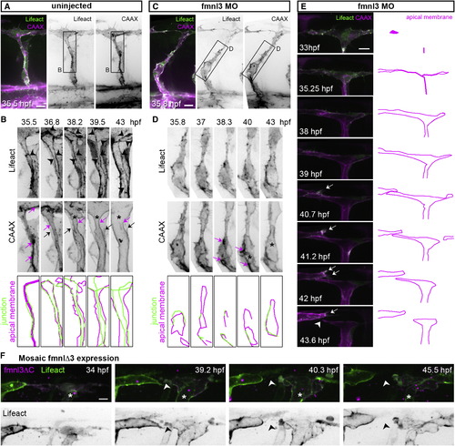
Junctional F-Actin Cables Assemble and Rearrange during Lumen Formation (A–D) Stills from time-lapse movies illustrating ISV lumenization in uninjected and in fmnl3 morpholino-injected Tg(fli1ep:Lifeact-EGFP);Tg(kdr-l:ras-Cherry)s916 embryos. The magenta arrow shows apical membrane. The black arrow show basal membrane, and the arrowhead shows junctional F-actin cable. The asterisk shows lumen in (B) and vacuole in (D). Scale bars represent 10 µm. (E and F) Stills from a time-lapse movie of the DLAV from a Tg(fli1ep:Lifeact-EGFP);Tg(kdr-l:ras-Cherry)s916 embryo injected with fmnl3 morpholino (E) or Tg(fli1ep:Lifeact-EGFP) embryo with mosaic fmnl3ΔC expression (*) (F). The arrow shows activated EC, and the arrowhead shows vessel disconnection. Scale bar represents 10 µm. See also Figure S3 and Movies S1, S2, and S3. |
Reprinted from Developmental Cell, 32, Phng, L.K., Gebala, V., Bentley, K., Philippides, A., Wacker, A., Mathivet, T., Sauteur, L., Stanchi, F., Belting, H.G., Affolter, M., Gerhardt, H., Formin-mediated actin polymerization at endothelial junctions is required for vessel lumen formation and stabilization, 123-32, Copyright (2015) with permission from Elsevier. Full text @ Dev. Cell