
|
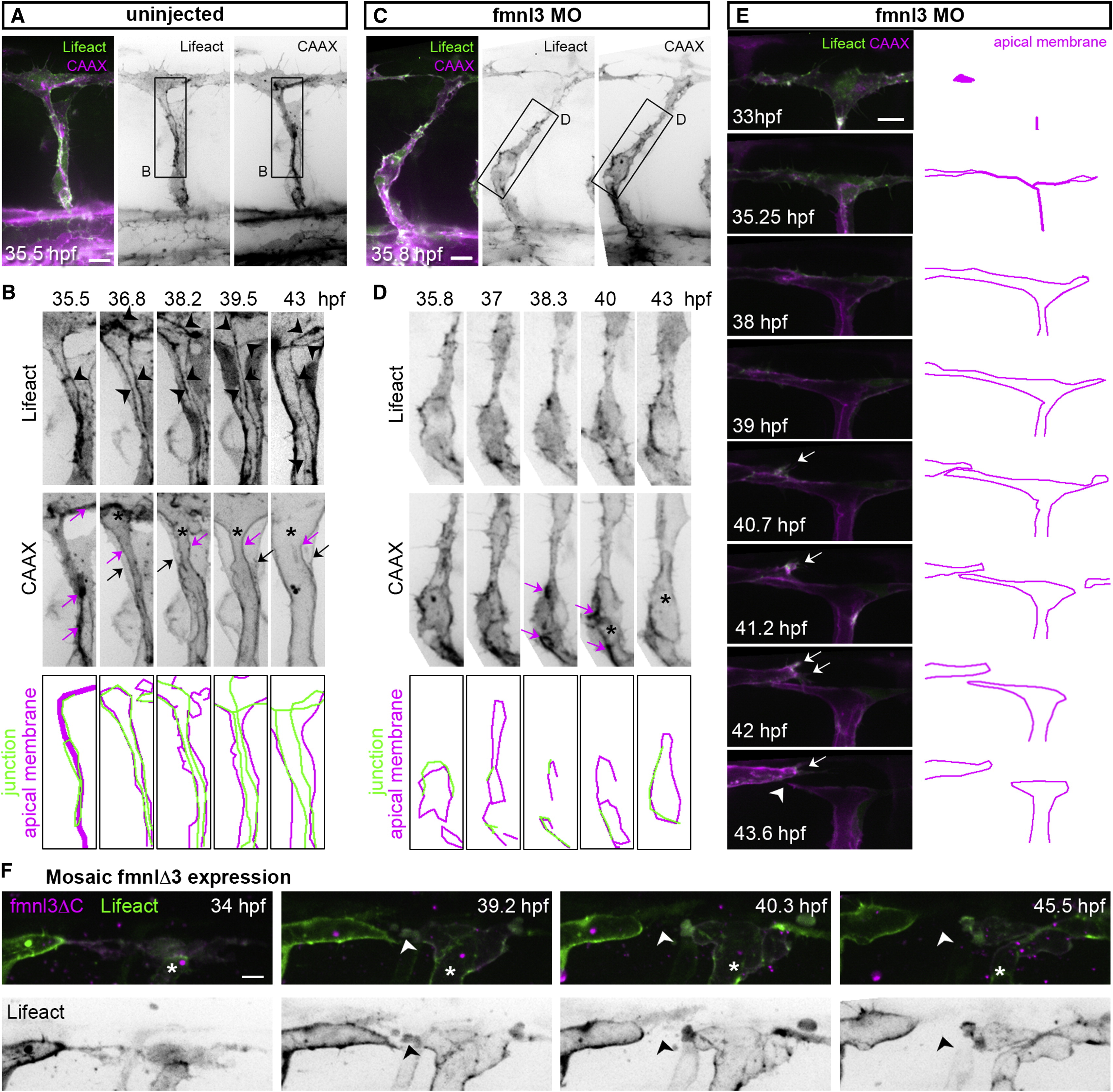
Fig. 3
Junctional F-Actin Cables Assemble and Rearrange during Lumen Formation
(A–D) Stills from time-lapse movies illustrating ISV lumenization in uninjected and in fmnl3 morpholino-injected Tg(fli1ep:Lifeact-EGFP);Tg(kdr-l:ras-Cherry)s916 embryos. The magenta arrow shows apical membrane. The black arrow show basal membrane, and the arrowhead shows junctional F-actin cable. The asterisk shows lumen in (B) and vacuole in (D). Scale bars represent 10 µm.
(E and F) Stills from a time-lapse movie of the DLAV from a Tg(fli1ep:Lifeact-EGFP);Tg(kdr-l:ras-Cherry)s916 embryo injected with fmnl3 morpholino (E) or Tg(fli1ep:Lifeact-EGFP) embryo with mosaic fmnl3ΔC expression (*) (F). The arrow shows activated EC, and the arrowhead shows vessel disconnection. Scale bar represents 10 µm.
See also Figure S3 and Movies S1, S2, and S3.
Reprinted from Developmental Cell, 32, Phng, L.K., Gebala, V., Bentley, K., Philippides, A., Wacker, A., Mathivet, T., Sauteur, L., Stanchi, F., Belting, H.G., Affolter, M., Gerhardt, H., Formin-mediated actin polymerization at endothelial junctions is required for vessel lumen formation and stabilization, 123-32, Copyright (2015) with permission from Elsevier. Full text @ Dev. Cell