
Fig. 8
- ID
- ZDB-FIG-150313-34
- Publication
- Huc-Brandt et al., 2014 - Zebrafish Prion Protein PrP2 Controls Collective Migration Process during Lateral Line Sensory System Development
- Other Figures
- All Figure Page
- Back to All Figure Page
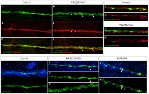
Loss of PLL nerve fasciculation and associated myelination in absence of PrP2. A–C. In control larvae at 5 dpf, sox10-GFP cells are tightly organized along the PLL nerve (nbt-dsred). D–F. Double morphant PrP2/p53 displayed an enlarged PLL nerve (with defasciculated axons, arrows) and rounded Schwann cells (arrowheads). G–G′. In 5 dpf larvae MBP labeling is observed in close apposition to sox10-GFP cells and formed a homogeneous line. H–H′. MBP labeling is altered and partially missing while Schwann cells are disorganized. I–I′. At 7 dpf, control larvae display tightly and regularly organized sox10-GFP positive cells. J–K′. In PrP2/p53 morphants (J–J′′), loosened Schwann cell processes are observed (arrows) as well as rounded cells (arrowheads) in PrP2 morphants (K–K′). Results obtained from five independent experiments (n = 160 embryos). |
| Genes: | |
|---|---|
| Fish: | |
| Knockdown Reagents: | |
| Anatomical Terms: | |
| Stage Range: | Day 5 to Days 7-13 |
| Fish: | |
|---|---|
| Knockdown Reagents: | |
| Observed In: | |
| Stage Range: | Day 5 to Days 7-13 |