
Fig. S6
- ID
- ZDB-FIG-150129-23
- Publication
- Zhao et al., 2014 - Leptin and IL-6 Family Cytokines Synergize to Stimulate Müller Glia Reprogramming and Retina Regeneration
- Other Figures
- All Figure Page
- Back to All Figure Page
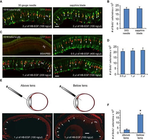
HB-EGF stimulates MG reprogramming and proliferation in the uninjured retina. Related to Figure 4. (A) A corneal puncture with a 30 gauge needle or a corneal incision made with a sapphire blade was followed by intravitreal injection of recombinant HB-EGF into the uninjured eye of 1016tuba1a:gfp fish. HB-EGF stimulated GFP expression and BrdU incorporation in MG throughout the retina’s inner nuclear layer. No GFP expression or BrdU incorporation is observed in PBS/BSA-injected eyes. (B) Quantification of BrdU+ cells in (A) shows similar effect of intravitreally delivered HB-EGF on MG proliferation when the cornea was either punctured with a needle or cut with a sapphire blade; n=3 per group. (C) A sapphire blade was used to make a small incision in the cornea and 200 ng of HB-EGF in 0.5 to 2 µl volumes was intravitreally injected into uninjured eyes of 1016tuba1a:gfp fish. (D) Quantification of BrdU+ cells following intravitreal delivery of different HB-EGF volumes reveals a similar effect on MG proliferation regardless of injection volume; n=3 per group. (E, F) HB-EGF must be delivered below the lens to stimulate MG proliferation; *P<0.05, n=3. Arrows point to MG-derived progenitor. Error bars, s.d. Scale bar, 50 µm (A, C); 150 µm (E). INL, inner nuclear layer. |