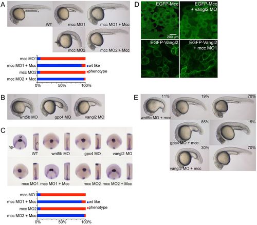
mcc acts downstream of wnt5b and vangl2 to regulate convergence and extension. (A) Injection of ATG (MO1) or splice junction (MO2) MOs targeting mcc yields zebrafish embryos with a shorter and ventrally curved anteroposterior axis with tightly packed somites at 1dpf. MO1 and MO2 positions are indicated in Fig. 1A. The black line with asterisk emphasizes the shortened yolk extension, and curved dotted line highlights the smaller eyes and reduced head development in mcc morphants compared with wild type (WT). The arrow indicates the region of anterior cell death. These defects are rescued by co-injection of mouse Mcc mRNA (see also supplementary material Fig. S2A). (B) Representative wnt5b, gpc4 and vangl2 morphants at 1dpf. (C) Triple ntl, hgg1 and dlx3 WISH at 10hpf. In wild-type embryos, hgg1 marks the polster (p) that lies on the anterior neural plate (np) boundary expressing dlx3. ntl1 identifies the thin midline notochord (inset). In morphants, the neural plate is wider mediolaterally, the notochord is thickened and shortened, and the polster is abnormally elongated posteriorly. Co-injection of Mcc mRNA and either mcc MO1 or MO2 yields hgg1/dlx3/ntl expression patterns that are indistinguishable from those of wild type (see also supplementary material Fig. S2B). (D) Lateral images of shield stage embryos injected as indicated. EGFP-Mcc is exclusively cytoplasmic in both wild-type and vangl2 morphant embryos. EGFP-Vangl2 is principally membrane localized in wild-type embryos, but is also visible in cytoplasmic puncta. EGFP-Vangl2 distribution is unaltered in mcc morphants. The N-terminal GFP-Xenopus laevis Mcc fusion (supplementary material Table S3) efficiently rescues zebrafish mcc morphants (data not shown). (E) Zebrafish mcc mRNA rescues loss of either wnt5b or vangl2 but not gpc4 at 1dpf. Combining mcc mRNA and wnt5b MO results in three phenotypic classes, as evidenced by variable yolk (and body axis) extension and head position. (A-E) MO and mRNA concentrations are provided in supplementary material Table S3. (A,C,E) Phenotypic distributions are indicated as percentages, with scored embryo counts listed in supplementary material Table S4.
|
