
Fig. 4
- ID
- ZDB-FIG-141120-4
- Publication
- van Leeuwen et al., 2014 - Modelling tuberculous meningitis in zebrafish using Mycobacterium marinum
- Other Figures
- All Figure Page
- Back to All Figure Page
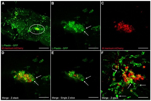
Composition of early granulomas. (A) Z-stack of the head of a zebrafish embryo (7 dpf) stained with anti-L-plastin, labeling phagocytic cells (green), infected with M. marinum E11 (red) via the caudal vein (5 dpi). Marked area is enlarged in panels B-E. (B-D) Z-stack of an early granuloma, with phagocytic cells stained with anti-L-plastin (B), M. marinum E11::mCherry (C) and merge (D). A phagocyte with high L-plastin intensity (closed arrow) and a phagocyte with low L-plastin intensity (open arrow) are depicted in panels B and D. (E) Single Z-slices of granuloma shows colocalization of phagocytes and mycobacteria. Arrows indicate different intensities of L-plastin, eliminating the possible effect of overlapping phagocytes in the Z-stack in panel B and D. (F) Z-stack of an early granuloma containing relatively more mycobacteria than phagocytes. Lots of phagocytes surrounding the cluster are probably still migrating to the cluster. The parenchymal infection route was used. Images were taken at 5 dpi. Scale bars: 150 µm (A), 35 µm (B-F). |