
Fig. 1
- ID
- ZDB-FIG-141120-1
- Publication
- van Leeuwen et al., 2014 - Modelling tuberculous meningitis in zebrafish using Mycobacterium marinum
- Other Figures
- All Figure Page
- Back to All Figure Page
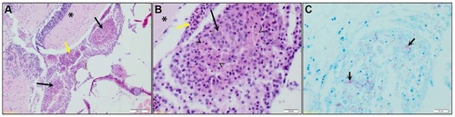
Granulomas in adult zebrafish after intraperitoneal infection. (A) Coronal section of adult zebrafish at 50 days after infection with M. marinum E11. Multifocal to coalescing granulomas (black arrows) affecting the meninges and submeningeal space structure can be seen, the brain parenchymal tissue (*) beneath the granulomas shows minimal lymphocytic inflammation, and the regional meningeal blood vessels are markedly congested (yellow arrow). Scale bar: 100 µm. Granuloma is enlarged in panels B and C. (B) Granulomas are composed of epithelioid and foamy macrophages (black arrow) that occasionally exhibited degeneration and necrosis (arrowheads). Scale bar: 20 µm. (C) Same granuloma as shown in panel B stained with ZN. Multiple acid-fast bacilli are present in the cytoplasm of the macrophages (black arrows). Scale bar: 20 µm. |