
Fig. 2
- ID
- ZDB-FIG-141027-1
- Publication
- Xu et al., 1994 - Spatially regulated expression of three receptor tyrosine kinase genes during gastrulation in the zebrafish
- Other Figures
- All Figure Page
- Back to All Figure Page
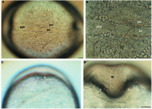
Axial hypoblast at the end of epiboly. Living zebrafish embryos at 9.5 h (A-C) and 11.5 h (D). Anterior is to the left in A and B; C and D are vegetal views looking at the head region. (A) Dorsal view of an embryo focused on the hypoblast. Presumptive notochord has segregated from paraxial mesoderm. (B) Axial hypoblast beneath the future hindbrain/midbrain. Anterior to the presumptive notochord, prechordal axial hypoblast continues as a strip towards the front of the embryo. (C,D) The ‘pillow’ of anterior hypoblast. At the end of epiboly there is a disclike accumulation of cells at the very anteriormost tip of the embryo (C). A few hours later, the forebrain has formed a depression in the anterior hypoblast giving the pillow its characteristic shape (D). Abbreviations: fb, forebrain; pn, presumptive notochord; pch, prechordal hypoblast; pm, paraxial mesoderm. Scale bars: 50 µm. |