
Fig. 2
- ID
- ZDB-FIG-141020-6
- Publication
- Fjose et al., 1994 - Expression of the zebrafish gene hlx-1 in the prechordal plate and during CNS development
- Other Figures
- All Figure Page
- Back to All Figure Page
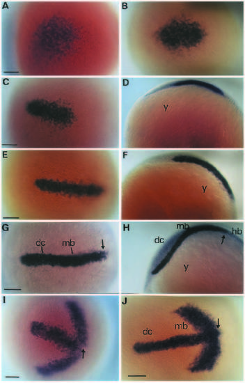
Expression of hlx-1 in the rostral region of zebrafish embryos during early stages of neurulation. Transcripts were localized by in situ hybridization on a series of whole-mount embryos at 9-10 hpf (A-H). Dorsal views of the rostral region (anterior to the left) of subsequent stages are shown in A,B,E and G. Side views of the same embryos as in C,E and G are shown in D,F and H, respectively. Some embryos were hybridized simultaneously with digoxigenin-labelled probes of both hlx-1 and the pax[b] gene (Krauss et al., 1991b). The double stained embryos shown in I and J are comparable to the stages presented in C and G, respectively. Arrows indicate the location of the presumptive midbrain-hindbrain boundary. Bar, 60 µm. Abbreviations: dc, diencephalon; hb, hindbrain; mb, midbrain; y, yolk. |
| Genes: | |
|---|---|
| Fish: | |
| Anatomical Terms: | |
| Stage: | 90%-epiboly |