
FIGURE
Fig. S4
- ID
- ZDB-FIG-141007-43
- Publication
- Palmyre et al., 2014 - Collective epithelial migration drives kidney repair after acute injury
- Other Figures
- All Figure Page
- Back to All Figure Page
Fig. S4
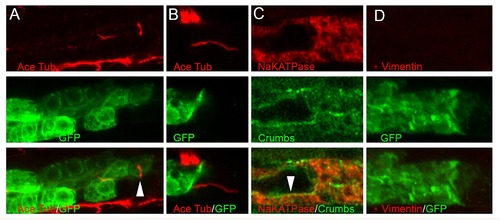
Epithelial and mesenchymal markers in injured epithelium. (A, B) Apical cilia at the edge of surviving epithelium. Upper panel – acetylated tubulin, middle panel – GFP, lower panel – combined. Panel (B) corresponds to figure 3B. (C) Higher magnification images corresponding to figure 3D. Upper panel – acetylated tubulin, middle panel – GFP, lower panel – combined. (D) Higher magnification images corresponding to figure 3F. Upper panel – acetylated tubulin, middle panel – GFP, lower panel – combined. |
Expression Data
Expression Detail
Antibody Labeling
Phenotype Data
Phenotype Detail
Acknowledgments
This image is the copyrighted work of the attributed author or publisher, and
ZFIN has permission only to display this image to its users.
Additional permissions should be obtained from the applicable author or publisher of the image.
Full text @ PLoS One