
Fig. 4
- ID
- ZDB-FIG-141007-42
- Publication
- Palmyre et al., 2014 - Collective epithelial migration drives kidney repair after acute injury
- Other Figures
- All Figure Page
- Back to All Figure Page
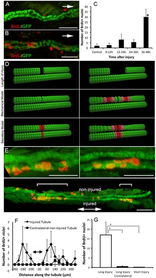
Kidney epithelial proliferation after acute injury. (A,B) Two examples of BrdU staining after segmental ablation early (0–12 hpi, A) and late (36–48 hpi, B) in ET11-9 GFP transgenic fish. Arrows indicate the direction of injury. Confocal slice thickness is 1.5 μm in (A), and 1.6 μm in (B). (C) Number of BrdU+ cells at various intervals after segmental laser ablation (0–12 hpi, n = 4, 12–24 hpi, n = 3, 24–36 hpi, n = 4, 36–48 hpi, n = 4) and compared to non-injured control condition (n = 8). There was no significant increase in cell proliferation early in the repair process (up to 36 hpi) but a pronounced increase in cell proliferation first detectable between 36 and 48 hpi, compared to control condition (p<0.05, C). (D) Two possible mechanisms that could trigger cell proliferation (mechanical stretch due to cell migration vs. a secretory factor) lead to different predictions about the distribution of cell proliferation after segmental ablation. Red color indicates the predicted distribution of cell proliferation after short (left panel) vs. long (right panel) tubule segment ablation. Upper row: initial injury; middle row: stretch response scenario (mechanical model). Lower row: secretory factor scenario. (E) Long segmental ablation resulted in two distinct bands of BrdU incorporation both upstream (left and lower sub-panels) and downstream (right and lower sub-panels, each sub-panel represents a confocal projection image). Brackets in the lower sub-panel indicate bands of proliferation upstream and downstream of the injury. The right and left upper sub-panels show a higher magnification of the areas of proliferation marked by brackets in the lower sub-panel. Other BrdU+ nuclei are outside of the kidney. (F) Comparison of BrdU incorporation in the injured (rhombi) vs. contralateral non-injured tubule (squares). This pattern of BrdU incorporation is most consistent with the mechanical model of the cell proliferation trigger. Double-arrow bar indicates the approximate site of injury. (G) Total number of BrdU+ nuclei after long (80–100 μm) vs. short (20 μm) segmental ablation (24–48 hpi). Long segmental ablation resulted in significantly increased number of BrdU+ cells compared to a contralateral non-injured side (as well as kidney epithelium after short injury or in a non-injured control). Long injury vs. contralateral non-injured tubule: p = 0.039, long injury vs. short injury: p = 0.048, n = 3 per each condition. Scale bars in (A,B and E upper sub-panels) = 30 μm, and 60 μm in (E, lower sub-panel). |