
Fig. 4
- ID
- ZDB-FIG-141006-18
- Publication
- Bernier et al., 2014 - Disruptive CHD8 Mutations Define a Subtype of Autism Early in Development
- Other Figures
- All Figure Page
- Back to All Figure Page
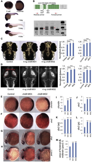
chd8 Disruption Results in Ectopic Expression of Forebrain/Midbrain Markers during Zebrafish Development In situ hybridization is shown for multiple markers before and after injection of chd8-MO1-4. (A) In situ hybridization of 2-cell, 5-somite, 1-, 3-, and 4-day stage zebrafish embryos using a 1.5 kbp chd8 antisense probe. chd8 is ubiquitously expressed in early stages; however, after day 1, its expression is enriched in the head region and the GI duct. (B) Two sets of morpholinos are independently designed to target two exon/intron junctions of chd8. To validate the morpholino effects, total mRNA was extracted at 24 hr postfertilization (hpf) followed by reverse transcription and PCR using primers flanking the targeted junctions. MO1 or MO2 injection leads to inclusion of the adjacent intron to mature mRNA. Red arrows indicate the PCR products of morpholino-modified chd8 transcripts. Uninjected AB strain embryos were used as control for all experiments in Figure 4. Data are represented as mean ± SEM. (C) The distance between the convex tips of the eyes was measured. MO1, MO2, MO3, and MO4 injection caused enlargement of this distance, and the results were quantified in the right panel. Data are represented as mean ± SEM. (D) Embryos were immunostained to highlight the neuronal axon tract in the developing brains. Embryos are imaged in dorsal view, and the optic tecta structure is indicated (red oval). The average distance between the optic tecta is measured and quantified (n = 50). Injection of chd8-MO1, MO2, MO3, and MO4 showed an enlargement of the distance consistent with interorbital distance measurements. Data are represented as mean ± SEM. (E and F) (E) Expression of chordin (marker of the dorsal organizing region) at shield stage, animal pole view, and dorsal oriented toward right. Chordin expression is expanded upon injection of chd8 morpholinos, and the overall staining intensity is quantified. (F) Orthodenticle homeobox 2 (otx2), an early marker of midbrain and forebrain neuronal progenitors. Expression of otx2 at tail bud stage, lateral view and dorsal oriented toward the right. Otx2 expression is enhanced upon injection of chd8 morpholinos and the overall staining intensity is quantified. (G and H) (G) Distal-less homeobox 2 (dlx2), a marker of neural stem cells. Expression of dlx2 at 24 hr stage, lateral view. Arrows point at the telencephalon (tel) region, and arrowhead points at the prethalamus (pt) region. dlx2 expression in the prethalamus, but not telencephalon, region is enhanced upon injection of chd8 morpholinos, as shown in (H) (pt) and (G) (tel), respectively. (H) Paired-box 2.1 (pax2.1), a marker of the midbrain/hindbrain boundary (MHB). Expression of pax2.1 at 24 hr stage, lateral view. Arrow points at MHB. The area of the head that contains the forebrain and midbrain is outlined by dashed red lines. The forebrain/midbrain region is enlarged upon injection of chd8 morpholinos, which is quantified. (I–M) Quantification of significant in situ hybridization results. n.s., not significant; p < 0.001; p < 0.0001. Data are represented as mean ± SEM. See also Figures S4, S5, and S6. |
| Genes: | |
|---|---|
| Antibody: | |
| Fish: | |
| Knockdown Reagents: | |
| Anatomical Terms: | |
| Stage Range: | 2-cell to Day 4 |
| Fish: | |
|---|---|
| Knockdown Reagents: | |
| Observed In: | |
| Stage: | Day 4 |
Reprinted from Cell, 158(2), Bernier, R., Golzio, C., Xiong, B., Stessman, H.A., Coe, B.P., Penn, O., Witherspoon, K., Gerdts, J., Baker, C., Vulto-van Silfhout, A.T., Schuurs-Hoeijmakers, J.H., Fichera, M., Bosco, P., Buono, S., Alberti, A., Failla, P., Peeters, H., Steyaert, J., Vissers, L.E., Francescatto, L., Mefford, H.C., Rosenfeld, J.A., Bakken, T., O'Roak, B.J., Pawlus, M., Moon, R., Shendure, J., Amaral, D.G., Lein, E., Rankin, J., Romano, C., de Vries, B.B., Katsanis, N., Eichler, E.E., Disruptive CHD8 Mutations Define a Subtype of Autism Early in Development, 263-76, Copyright (2014) with permission from Elsevier. Full text @ Cell