
Fig. 7
- ID
- ZDB-FIG-140811-43
- Publication
- Zhu et al., 2014 - Haploinsufficiency of Def Activates p53-Dependent TGFbeta Signalling and Causes Scar Formation after Partial Hepatectomy
- Other Figures
- All Figure Page
- Back to All Figure Page
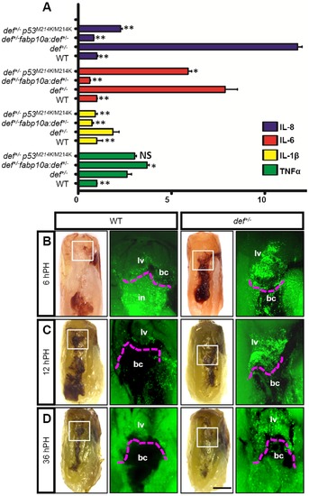
def+/- mutant liver exhibited a prolonged inflammatory reaction at the amputation site after PH. (A) qPCR analysis of cytokine genes TNFα, IL-1β, IL-6 and IL-8 in adult wild-type, def+/-, fabp10a:def+/- and def+/- p53M214K/M214K livers prior to the surgical procedure. Gene expression is expressed as the fold change after normalisation against β-actin. The value for each gene in the wild-type was set at 1. The p value represents the statistical differences between def+/- and another corresponding group. *: p<0.05, **: p<0.01, NS: not significant. (B–D) Adult fish of Tg(zlyz:EGFP) in a wild-type and def+/- background were subjected to PH, and collected 6 h after PH (B), 12 h after PH (C) and 36 h after PH (D) for EGFP fluorescence imaging. Images were taken from the region outlined in white in the corresponding bright field picture shown on the left. The region above the dashed magenta lines is the liver tissue adjacent to the amputation site. Black asterisks highlight the EGFP signal yielded possibly from neutrophils in the intestine. A representative image is shown for each time-point. bc, blood clot; in, intestine; lv, liver tissue. Scale bar: 5 mm (bright field) and 2 mm (GFP field). |
| Gene: | |
|---|---|
| Fish: | |
| Condition: | |
| Anatomical Term: | |
| Stage: | Adult |
| Fish: | |
|---|---|
| Condition: | |
| Observed In: | |
| Stage: | Adult |