
Fig. 3
- ID
- ZDB-FIG-140811-39
- Publication
- Zhu et al., 2014 - Haploinsufficiency of Def Activates p53-Dependent TGFbeta Signalling and Causes Scar Formation after Partial Hepatectomy
- Other Figures
- All Figure Page
- Back to All Figure Page
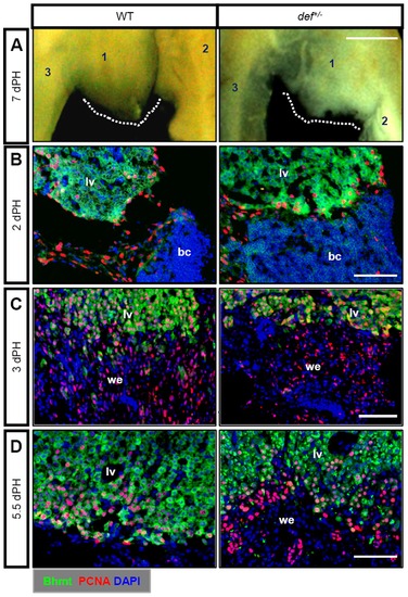
Def haploinsufficiency results in a failure in the remodelling of the wound epidermis to liver tissue. (A) Ventral view of the gross morphology of an amputated adult wild-type and def+/-liver 7 days after PH. The dotted white line indicates the amputation site. 1: ventral lobe, 2: left dorsal lobe, 3: right dorsal lobe. (B–D) Representative images of co-immunostaining of PCNA and Bhmt to compare the process of epithelialisation of the amputation site 2 days after PH (B), wound epidermis formation 3 days after PH (C), and wound epidermis remodelling 5.5 days after PH (D) between the wild-type and the def+/- mutant. Nuclei were stained with 4′,6-diamidino-2-phenylindole (DAPI) (blue). In each case or at each time-point, more than 10 sections from at least three wild-type or def+/- mutant fish were examined. bc, blood clot; lv, liver tissue; we, wound epidermis. Scale bar: 5 mm (A) and 75 μm (B–D). |
| Antibody: | |
|---|---|
| Fish: | |
| Condition: | |
| Anatomical Term: | |
| Stage: | Adult |
| Fish: | |
|---|---|
| Condition: | |
| Observed In: | |
| Stage: | Adult |