|
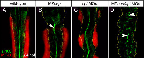
Lack of mesoderm disrupts neural tube morphogenesis in both anterior and posterior levels of the neuro-axis. (A,B) Projection of confocal z-series showing dorsal view of mesoderm (red), expressing the muscle marker (MF-20) and aPKC (green) in caudal hindbrain and anterior spinal cord at 24 hpf. The normal midline domain of aPKC is maintained in regions of MZoep embryo that are adjacent to mesoderm (arrowhead). (C) Mesoderm is still present but disrupted in spt-deficient embryos and the aPKC domain is largely normal. (D) Embryos deficient for both oep and spt have no mesoderm adjacent to the spinal cord and apical aPKC is severely disrupted in spinal cord (arrowheads). Basal extremity of neural tube indicated by yellow dotted lines. aPKC, atypical protein kinase C; hpf, hours post fertilization; MZoep, maternal-zygotic one-eyed pinhead.
|