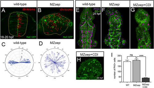
Ectopic divisions do not generate the abnormal neural tube in MZoep embryos. (A) Location and orientation of divisions monitored over a 2-hour time-lapse period of neural rod development in wild-type embryo by analyzing the expression of apical marker Pard3-GFP (green) and H2B-RFP mRNA to label nuclei (not included in image for clarity, red dumbbells indicate location and orientation of dividing cells). Wild-type neural progenitor divisions are strongly orientated along the mediolateral axis of the developing neural tube. Yellow dots outline the rod. (B) Location and orientation of divisions monitored over a 2-hour time-lapse period of neural rod development in MZoep embryo. (C,D) Orientation plots of divisions in wild-type and MZoep embryos. (E,F,G) Dorsal projections of ventricle morphology (ZO-1, green) at 24 hpf in wild-type, MZoep embryos and MZoep embryos treated with CDIs (hydroxyurea and aphidicolin). Blocking division does not rescue ventricle morphology. PH3 staining of mitotic figures (purple) used to calculate efficiency of division block. (H) Transverse section of brain from MZoep embryo treated with division blockers. (I) Quantification of divisions in wild-type, MZoep and MZoep division blocked embryos. Number of cell divisions between wild-type 194 and MZoep 206, P = 0.5129 Student’s t-test and between MZoep 206 and MZoep + CDI 39, ***P <0.0001 Student’s t-test. In (E), (F) and (G), equivalent tissue volumes were used (360 µm in length x 130 µm in width x 90 µm in depth). In (I), error bars indicate SEM. CDI, cell division inhibitor; H2B-RFP, histone H2B/red fluorescent protein fusion; hpf, hours post fertilization; MZoep, maternal-zygotic one-eyed pinhead; Pard3-GFP, green fluorescent protein/polarity protein partitioning defective 3 fusion; PH3, phospho-histone 3 marker; SEM, standard error of the mean; wt, wild-type; ZO-1, zonula occludens 1.
|
