|
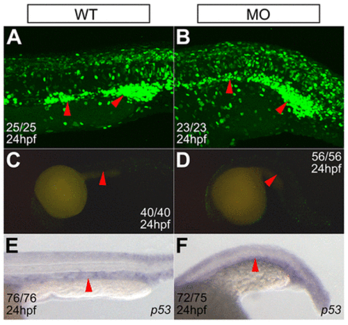
ptpn9a knockdown causes no defect in hematopoietic cell proliferation and apoptosis. (A,B) ptpn9a-MOs were injected into Tg(ef1α:mAG-zGem) transgenic embryos, in which green-fluorescence-positive cells are in phases S, G2 or M of the cell cycle. Note that there is little difference in the numbers of green-fluorescence-positive cells in the ICM between ptpn9a-morphant (B) and wild-type (A) embryos at 24 hpf. (C,D) The TUNEL assay showed comparable numbers of apoptotic cells in the ICM in ptpn9a morphants (D) and controls (C) at 24 hpf. (E,F) RNA in situ hybridization revealed that expression of the p53 gene was not induced in the ICM of ptpn9a-morphant embryos (F), compared with wild-type controls (E). Red arrowheads indicate the ICM. The number of phenotypic embryos/total embryos is indicated.
|