
Fig. 2
- ID
- ZDB-FIG-140523-13
- Publication
- Van Ryswyk et al., 2014 - The role of inab in axon morphology of an identified zebrafish motoneuron
- Other Figures
- All Figure Page
- Back to All Figure Page
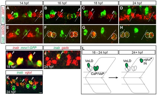
inab is dynamically expressed in PMNs and INs. (A–K) Single confocal slices of embryos labeled with inab, islet, and neurotransmitter riboprobes (gads for GABA and vglut for glutamate). Diagonal lines represent somite boundaries. At 14 hpf, inab is coexpressed with neither islet1 (A) nor islet2a (E). Between 16 hpf and 24 hpf, inab is expressed in islet2a+ PMNs (circled cells in F–H) but not islet1+ PMNs (arrowheads in A–C). inab is expressed in the VeLD IN, as determined by coexpression with both GFP in mnx1:GFP transgenic embryos (arrowheads, I) and gad mRNA (arrowhead, J). At 24 hpf, inab is coexpressed with vglut (arrowheads, K) in a cell dorsal to the VeLD IN (circle, K). (L) Schematic of inab mRNA dynamics during early development. Between 16–24 hpf, inab is expressed in both CaP and VaP MNs and in VeLD INs. After 24 hpf, inab expression in CaP and VaP is downregulated, although it persists in VeLD and an additional, dorsally-located, glutamatergic IN. Scale bar, 5 μm in A–J; 10 μm in K. |
| Genes: | |
|---|---|
| Fish: | |
| Anatomical Terms: | |
| Stage Range: | 10-13 somites to Prim-5 |