
Fig. 1
- ID
- ZDB-FIG-140515-10
- Publication
- Toruno et al., 2014 - Interdependence of Bad and Puma during ionizing-radiation-induced apoptosis
- Other Figures
- All Figure Page
- Back to All Figure Page
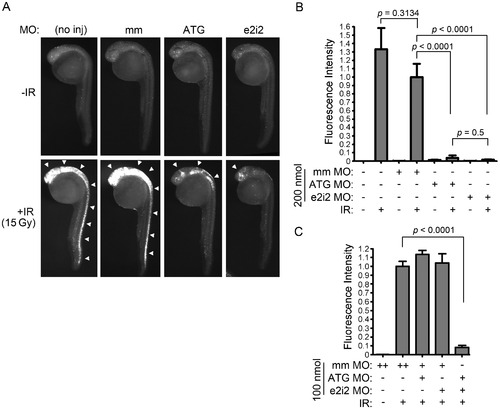
Bad is required for IR-induced apoptosis in zebrafish embryonic neural tissue. (A) Shown are lateral views of 27-hpf embryos (head is top left in each panel) either uninjected or injected with 200 nmol of bad ATG, bad e2i2 or mismatch (mm) MO. Half of each group of embryos were exposed to 15 Gy IR, and all were analyzed by the Casp3 assay. In control embryos (no inj and mm), IR-induced apoptosis occurs predominantly in the brain and all along the spinal cord (white arrowheads), whereas in bad-deficient embryos (ATG and e2i2), residual apoptosis is only observed in the head (arrowheads). (B) Fluorescence intensity, reflecting level of Caspase 3 activity, was measured in the spinal cords of at least 10 embryos from each group in (A) as previously described [34]. The fluorescence intensity in irradiated mismatch-MO-injected embryos was normalized to 1. (C) One-cell stage zebrafish embryos were injected with 100 nmol of bad ATG, bad e2i2 or mm MO as indicated (“++” indicates that 200 nmol was injected to keep total concentration of MO constant between experimental groups) and irradiated and analyzed as in (A-B). Data represent one experiment, and the experiment was independently performed three times with similar results. |
| Antibody: | |
|---|---|
| Fish: | |
| Condition: | |
| Knockdown Reagents: | |
| Anatomical Terms: | |
| Stage: | Prim-5 |
| Fish: | |
|---|---|
| Condition: | |
| Knockdown Reagents: | |
| Observed In: | |
| Stage: | Prim-5 |