
FIGURE
Fig. S5
Fig. S5
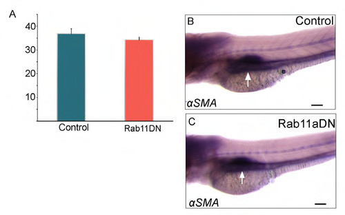
Cell number and mesenchymal differentiation is not impaired in Rab11DN embryos. (A) Quantification of total cell number in the gut in WT and Rab11aDN embryos. WT n=13, DN n=11, P>0.32. (B,C) Lateral view of an in situ hybridization showing αSMA expression in WT and Rab11aDN embryos at 72 hpf. Arrow points to smooth muscle. Scale bar: 100 μm. (D-E′′) Confocal section of WT and Rab11DN embryo stained for Myh11. Arrowhead points to Mhy11 in the mesenchyme. Asterisk indicates non-specific epithelial staining as observed previously (Wallace et al., 2005). Scale bar: 20 μm. |
Expression Data
Expression Detail
Antibody Labeling
Phenotype Data
Phenotype Detail
Acknowledgments
This image is the copyrighted work of the attributed author or publisher, and
ZFIN has permission only to display this image to its users.
Additional permissions should be obtained from the applicable author or publisher of the image.
Full text @ Development