
Fig. 3
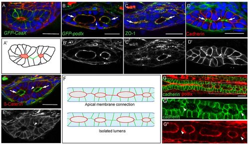
Cell-cell contacts are found between lumens. (A-E) Confocal cross sections of embryos at the lumen resolution stage. (A) Tg(hsp70l:GFP-CaaX) labels all cell membranes. (A2) Cartoon diagram of Fig. 3A depicting laterally arranged lumens in red and ‘bridge’ contacts in green. (B,B2) Apical protein, GFP-Podocalyxin surrounds the lumen but is not found at bridge contacts. Phalloidin (red). (C,C2) Antibody staining against Zo-1 labels tight junctions. Phalloidin (red). (D-E2) Antibody staining against cadherin and β-catenin labels basolateral contacts and ‘bridge’ contacts between lumens. Phalloidin (green). (F) Cartoon depicting two scenarios of lumen fusion along the AP axis. Apical membrane (red) can be deposited on membranes between lumens (top) or lumens may arise isolated and fuse directly without an apical membrane linker (bottom). (G-G2) Whole-mount confocal image of a lumen resolution stage embryo expressing GFP-Podocalyxin (red) and stained for cadherin in green. Cadherin localizes to basolateral contacts separating lumens. Arrows indicate lumens; arrowheads indicate bridge contact. Scale bars: 20 μm. |
| Genes: | |
|---|---|
| Antibodies: | |
| Fish: | |
| Condition: | |
| Anatomical Terms: | |
| Stage: | Long-pec |