
Fig. 4
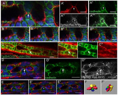
Basolateral adhesions separate lumens along the AP axis. (A-A22) Whole-mount confocal image of an embryo expressing GFP-Podocalyxin (false colored in red) and stained for cadherin (green) shows luminal expansion during a fusion event. Ras-RFP (white) marks cell membranes. Arrows mark fusion event. Blue, DAPI. (B-B22) Optical sections from a z-stack surrounding a fusion event. (C-C2) Whole-mount confocal image of a TgBAC(GFP-cldn15la) embryo shows a putative adhesion snapping event during fusion. The arrow marks adhesion at the surface. (D-D2) Whole-mount confocal image of an embryo expressing GFP-Podocalyxin (red) and stained for cadherin (green) shows adhesion snapping during fusion. Ras-RFP (white) marks cell membranes. The arrow marks adhesion at the surface. (E-E2) Optical sections from z-stack surrounding a fusion event. The asterisk marks a cell with adhesion. (F,F2) Space-fill projection labeling cells surrounding the fusion event. Lumen, red. The asterisk marks a cell with adhesion. Scale bars: 10 μm. |
| Gene: | |
|---|---|
| Antibody: | |
| Fish: | |
| Condition: | |
| Anatomical Terms: | |
| Stage: | Long-pec |