
FIGURE
Fig. S8
- ID
- ZDB-FIG-140507-4
- Publication
- Stewart et al., 2014 - Sequential and opposing activities of Wnt and BMP coordinate zebrafish bone regeneration
- Other Figures
- All Figure Page
- Back to All Figure Page
Fig. S8
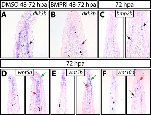
Localization of Wnt and BMP signals during fin regeneration. (A and B) In situ hybridization using a dkk3b probe on 72 hpa fins from control (A, DMSO 48- 72 hpa) and BMPRi (B, 5 μM, 48-72 hpa) treated fish. (C-F) Expression of bmp2b (C), wnt5a (D), wnt5b (E), and wnt10a (F) by in situ hybridization on paraffin sections from 72 hpa fin, shown at low and high magnification. The black arrows indicate osteoblasts, red arrows point to distal blastema mesenchymal cells, and green arrows mark basal epidermal cells. |
Expression Data
Expression Detail
Antibody Labeling
Phenotype Data
Phenotype Detail
Acknowledgments
This image is the copyrighted work of the attributed author or publisher, and
ZFIN has permission only to display this image to its users.
Additional permissions should be obtained from the applicable author or publisher of the image.
Full text @ Cell Rep.