
Fig. S2
- ID
- ZDB-FIG-140428-30
- Publication
- Graciarena et al., 2014 - Dynamics of axonal regeneration in adult and aging zebrafish reveal the promoting effect of a first lesion
- Other Figures
- All Figure Page
- Back to All Figure Page
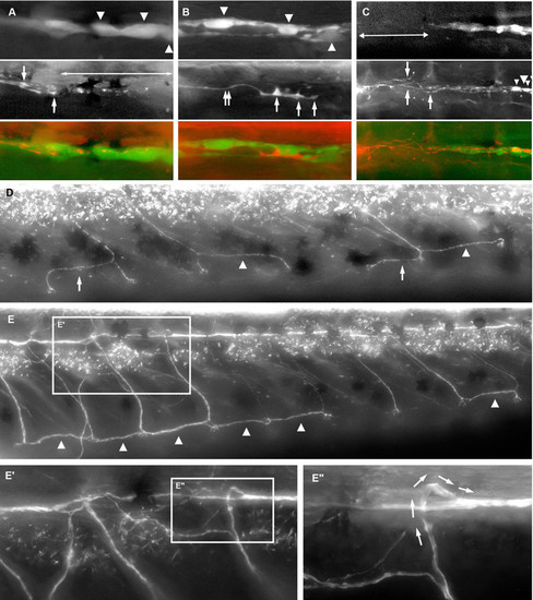
Involvement of Schwann cells in axonal regeneration. (A) Three days after nerve cut in 1-mo postfertilization fishes, the axons have fully degenerated (double-headed arrow), but Schwann cells (arrowheads) are not noticeably affected posterior to the leading growth cones (arrows). (B) During regeneration, the leading growth cone (arrows) and neurite (double arrow) are closely apposed to Schwann cells (arrowheads). (C) 2 d after nerve ablation, Schwann cells are still absent from the exposed region (double-headed arrow), yet the growth cones of regenerating neurites (arrowheads) have crossed the glial gap. Regenerating neurites are notably defasciculated over the ablated region (arrows). (D) Reinnervation may proceed from one neuromast to the next, either anteriorly (arrows) or posteriorly (arrowheads). (E-E3) Regenerating axons may grow ventrally and then dorsally again to join the major nerve path (arrows in E3). |