
Fig. 1
- ID
- ZDB-FIG-140428-26
- Publication
- Graciarena et al., 2014 - Dynamics of axonal regeneration in adult and aging zebrafish reveal the promoting effect of a first lesion
- Other Figures
- All Figure Page
- Back to All Figure Page
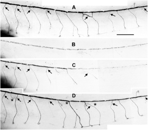
Patterns of PLL nerve regeneration. (A–D) Nerve regeneration in a mosaic fish in which most PLL neurons are labeled. Nerve branches on somites 14–25 before the nerve was cut (A), 2 d later (B), 3 d later (C), and 6 d later (D). Wallerian degeneration is evident in B and C. Regeneration has reached somite 13, just left of panel B, after 2 d and was complete including the caudal lines after 6 d. Arrows in A point to slight departures from the usual pattern; all departures were faithfully reproduced by the regenerating axons (arrows in D). Asterisks in D point to differences between the original and the regenerated patterns. The cut was done at the level of somite 2 on a 1.5-mpf mosaic fish derived from a wild-type blastula transplanted with cells from a nbt:dsred donor blastula. Most or all PLL neurons were fluorescent on the left side of this particular mosaic fish. (Scale bar, 500 μm.) |