
Fig. 5
- ID
- ZDB-FIG-140422-43
- Publication
- Holly et al., 2014 - Sfrp1a and Sfrp5 function as positive regulators of Wnt and BMP signaling during early retinal development
- Other Figures
- All Figure Page
- Back to All Figure Page
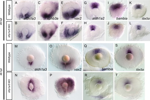
sfrp1a/sfrp5 regulate retinal dorso-ventral patterning. To examine dorso-ventral retina patterning in sfrp1a/5 MO co-injected embryos, we employed in situ hybridization of ventral marker genes aldh1a3, ephb3a and vax2, as well as dorsal markers aldh1a2, bambia, and tbx5a at 28 and 48 hpf. At 28 hpf, sfrp1a/5 MO injected embryos display a normal domain of ventral markers compared to WT, though with slightly reduced levels (A–F). Analysis of 28 hpf dorsal retina patterning in sfrp1a/5 MO embryos also demonstrates a decreased expression of markers (G–L). By 48 hpf, in comparison to controls, sfrp1a/5 MO injected embryos display an expansion of both aldh1a3 and vax2 expression into the dorsal regions of the retina (M–P). Analysis at 48 hpf demonstrates a clear reduction of tbx5a and bambia expression domains and levels in sfrp1a/5 MO injected embryos (Q–T). |
| Genes: | |
|---|---|
| Fish: | |
| Knockdown Reagents: | |
| Anatomical Terms: | |
| Stage Range: | Prim-5 to Long-pec |
Reprinted from Developmental Biology, 388(2), Holly, V.L., Widen, S.A., Famulski, J.K., and Waskiewicz, A.J., Sfrp1a and Sfrp5 function as positive regulators of Wnt and BMP signaling during early retinal development, 192-204, Copyright (2014) with permission from Elsevier. Full text @ Dev. Biol.