
FIGURE
Fig. 1
- ID
- ZDB-FIG-140422-10
- Publication
- Yamanaka et al., 2014 - In vitro analysis suggests that difference in cell movement during direct interaction can generate various pigment patterns in vivo
- Other Figures
- All Figure Page
- Back to All Figure Page
Fig. 1
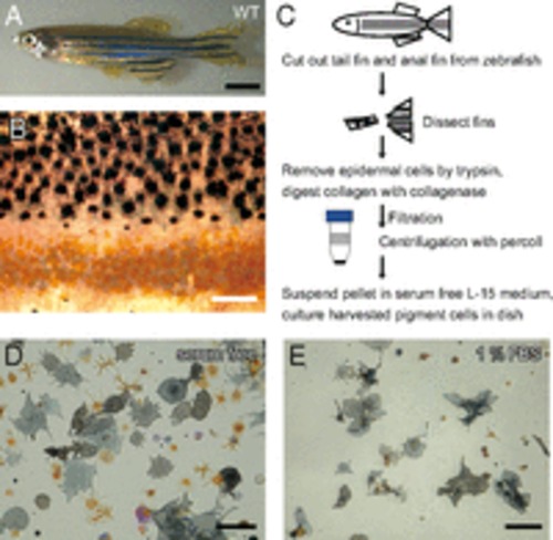
Harvesting pigment cells from zebrafish fins. (A) Adult WT zebrafish. (B) Magnified image of the surface of a WT fish body. Black spots are melanophores, and yellow spots are xanthophores. Clear gaps exist between the regions containing melanophores and the regions containing xanthophores. (C) A schematic diagram describing the harvest of pigment cells from fins. (D and E) The effect of serum in the medium on cell spreading. Cell pellets were suspended in (D) serum-free L15 medium or (E) medium containing 1% FBS. (Scale bars: A, 5.0 mm; B, D, and E, 100 μm.) |
Expression Data
Expression Detail
Antibody Labeling
Phenotype Data
Phenotype Detail
Acknowledgments
This image is the copyrighted work of the attributed author or publisher, and
ZFIN has permission only to display this image to its users.
Additional permissions should be obtained from the applicable author or publisher of the image.
Full text @ Proc. Natl. Acad. Sci. USA