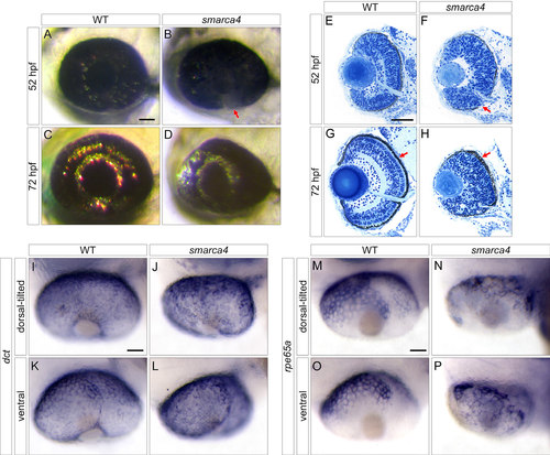
Smarca4 RPE development is abnormal. A-D: Whole-mount wild type (WT) and SWI/SNF-related, matrix associated, actin dependent regulator of chromatin, subfamily a, member 4 (smarca4) eyes at 52 and 72 h post-fertilization (hpf) are shown. The lateral view is shown. Anterior is to the left, and dorsal is up. The red arrow in B indicates a ventral region of smarca4 where the retinal pigment epithelium (RPE) pigmentation level was lower than the other regions. E–H: Transverse histological sections of the WT and smarca4 eyes at 52 and 72 hpf are shown. One distinctive hallmark of smarca4 is the disruption of their retinal lamination. In addition, the smarca4 RPE appears paler (B and E, red arrow) and dysmorphic (H, red arrow) compared with the WT. In these sections, lateral is to the left, and dorsal is up. The RPE differentiation problem was further illustrated with in situ hybridization with dct (I-L) and rpe65a (M-P) at 52 hpf. The expression of dct was comparable between WT and smarca4 RPE, suggesting that the RPE cells in smarca4 were still committed to the pigment cell lineage. Nonetheless, the differentiation of smarca4 was abnormal. This was demonstrated by the slightly irregular staining of dct and very irregular staining of rpe65a, a differentiation marker. In both cases, the dorsal-tilted view (I, J, M, and N) and the ventral view (K, L, O, and P) are shown, and the anterior side is to the left. Scale bars=50 µm.
|
