
FIGURE
Fig. 5
- ID
- ZDB-FIG-140325-31
- Publication
- Hong et al., 2013 - Cholinergic left-right asymmetry in the habenulo-interpeduncular pathway
- Other Figures
- All Figure Page
- Back to All Figure Page
Fig. 5
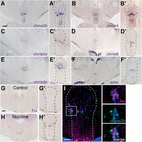
Nicotine-responsive neurons localize to iIPN. Transverse sections of adult brain show chrna2b (A and A2), chrnb4 (B and B2), chrna2a (C and C2), chrna5 (D and D2), chrnb2b (E and E2), and ache (F and F2) transcripts. Control (G and G2) and nicotine-treated (H and H2) fish were processed for fos expression. (Scale bars: A and G, 100 μm.) (A2–H2) Enlarged images of the IPN. (I) Double fluorescent ISH shows fos (magenta) and chrna2b (green) colocalization. DAPI labeling is in blue. The white box in I corresponds to enlarged panels on the right. (Scale bar: 50 μm.) Dotted lines delineate the IPN. |
Expression Data
| Genes: | |
|---|---|
| Fish: | |
| Anatomical Term: | |
| Stage: | Adult |
Expression Detail
Antibody Labeling
Phenotype Data
Phenotype Detail
Acknowledgments
This image is the copyrighted work of the attributed author or publisher, and
ZFIN has permission only to display this image to its users.
Additional permissions should be obtained from the applicable author or publisher of the image.
Full text @ Proc. Natl. Acad. Sci. USA