
Fig. 3
- ID
- ZDB-FIG-140325-29
- Publication
- Hong et al., 2013 - Cholinergic left-right asymmetry in the habenulo-interpeduncular pathway
- Other Figures
- All Figure Page
- Back to All Figure Page
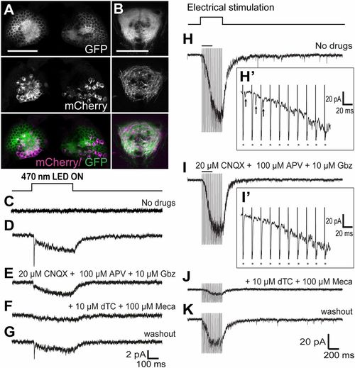
Sustained Hb stimulation induces slow cholinergic current in the vIPN. Confocal images of the dHb (A) and IPN (B) of 5-dpf larvae carrying the TgBAC(gng8:nfsB-CAAX-GFP)c375, TgBAC(gng8:GAL4)c426, and Tg(UAS:ChR2-mCherry)s1985t/+ transgenes. (Scale bars: 50 μm.) Averages of 15 responses evoked in a vIPN neuron by optogenetic stimulation (400 ms) of Hb neurons in control (C) and ChR2-mCherry brains in the absence of drugs (D), presence of 20 μM 6-cyano-7-nitroquinoxaline-2,3-dione (CNQX) + 100 µM amino-5-phosphonopentanoic acid (APV) + 10 μM gabazine (Gbz) (E), with addition of 10 μM (+)-d-tubocurarine chloride (dTc) + 100 μM mecamylamine (Meca) (F), and after washout (G). Examples of responses evoked in a single vIPN neuron by repetitive electrical stimulation of the right Hb (400 ms, 50 Hz) without drugs (H and H2), with 20 μM CNQX + 100 μM APV + 10 μM Gbz (I and I2), with addition of 10 μM dTc + 100 μM Meca (J), and after washout (K). H2 and I2 are higher resolution traces from H and I, respectively, illustrating fast EPSCs (H2, arrows). Asterisks in H2 and I2 mark artifacts of electrical stimulation. |
| Genes: | |
|---|---|
| Fish: | |
| Anatomical Terms: | |
| Stage: | Day 5 |