
Fig. 5
- ID
- ZDB-FIG-140321-12
- Publication
- Miller et al., 2000 - sucker encodes a zebrafish Endothelin-1 required for ventral pharyngeal arch development
- Other Figures
- All Figure Page
- Back to All Figure Page
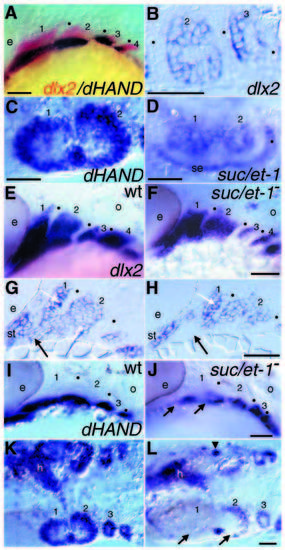
dlx2, dHAND, and suc/et-1 expression in wild-type (A-E,G,I,K) and suc/et-1 mutants (F,H,J,L). In all panels, arches are numbered and when visible, the pharyngeal pouches are marked with black dots. (A) Two-color in situ hybridization with dlx2 in red and dHAND in dark blue in a wild-type embryo at 28 hours. At this stage, dHAND expression marks a ventral subset of dlx2-expressing mesenchymal cells in each of the first four arches. (B) dlx2 expression in horizontal section through arches 2 and 3 at 28 hours. Within each arch, dlx2-expressing cells surround a core of nonexpressing cells. (C) Ventral view of dHAND expression in wholemount embryo at 28 hours. Within each arch, dHAND-expressing cells also surround a central core of non-expressing cells. (D) Ventral view at 28 hours showing suc/et-1 expression, which is strikingly complementary to dHAND expression (compare with C). suc/et-1 expression is detected in a central core of mesenchymal cells in the first two arches, and in arch epithelia, both ventral surface ectoderm and the second pharyngeal pouch. (E-F), Lateral views, at 28 hours, of dlx2 expression in PCR-genotyped wild-type (E) and suc/et-1 mutant (F) whole-mount embryos. In wild types dlx2 is expressed broadly in most if not all postmigratory arch neural crest, and is not expressed in the pharyngeal pouches. suc/et-1 mutant embryos have a reduction of ventral dlx2 expression. (G-H) 28 hours; sagittal sections of dlx2 expression in PCR-genotyped wild-type (G) and suc/et-1 mutant (H) cut through the lateral aspect of arches 1 and 2. In suc/et-1 mutant embryos, ventral cells are present but do not express dlx2 (black arrows). In contrast, suc/et-1 mutant dorsal cells (white arrows) and cells lining the stomodeum express dlx2 at normal levels. (I-L) dHAND expression in whole-mount wild-type (I, K) and suc/et-1 mutant (J,L) embryos at 28 hours. I and J are lateral views, K and L are ventral views. dHAND expression in suc/et-1 mutants is severely reduced in the first two arches (arrows in J and L). dHAND is also normally expressed in the heart, the pectoral fin (data not shown), and a discrete cluster of cells at the arch one/two boundary (arrowhead in L); these domains are not affected in suc/et-1 mutants (K-L and data not shown). e, eye; h, heart; o, otic vesicle; se, surface ectoderm; st, stomodeum. Scale bars: 50 μm. |